How White Blood Cell DNA Powers Cancer Blood Screening
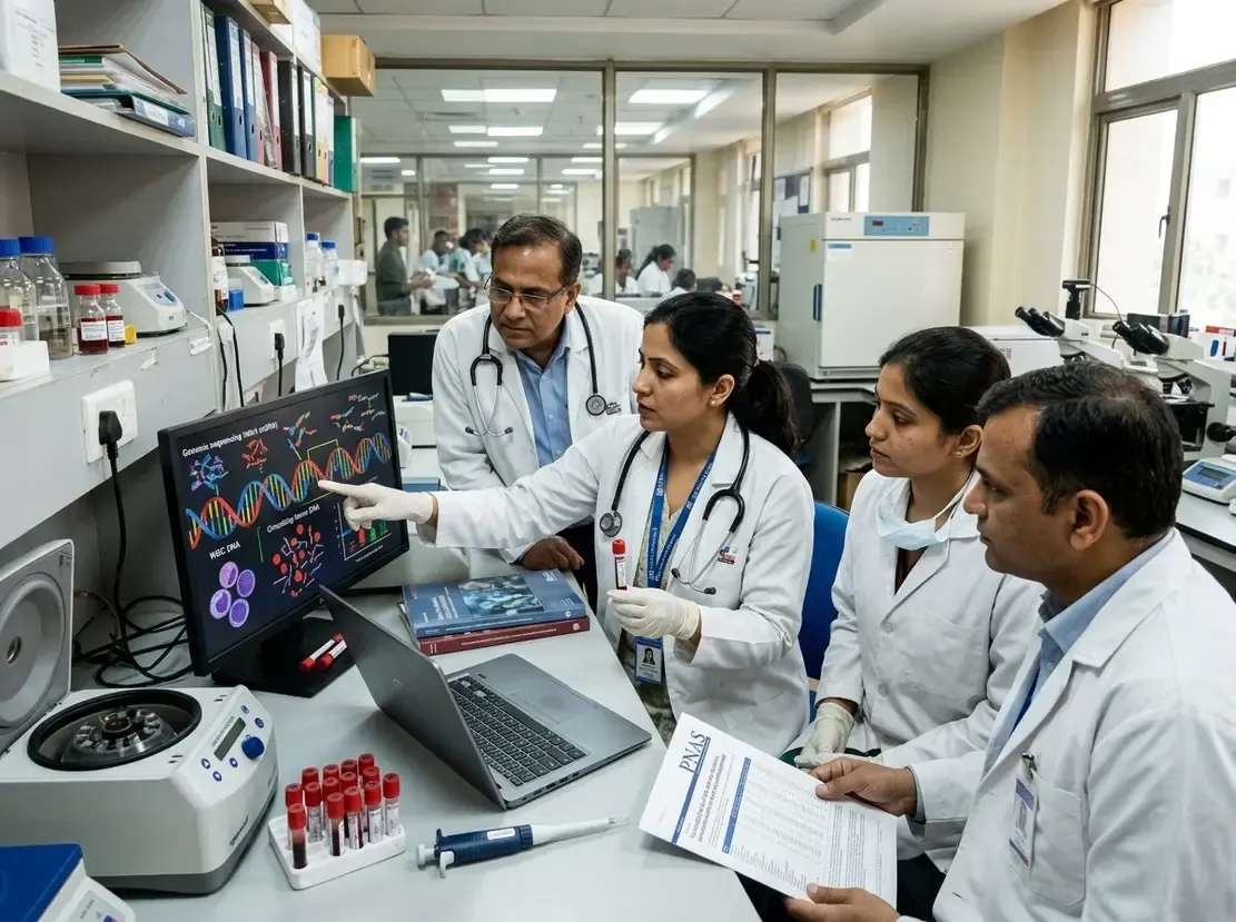
Recent clinical research from India has fundamentally challenged an established paradigm in blood-based cancer detection. Historically, oncologists assumed that genomic material shed by normal immune cells diluted tumor signals. However, a landmark study demonstrates that immune cell DNA correlates with stronger tumor signals in plasma.
Clinical Findings on White Blood Cell DNA Dynamics
The multicenter EMERGE study evaluated CancerSpot, an innovative multi-cancer early detection test developed by Strand Life Sciences. Over four years, investigators examined blood samples from 1,480 participants across 43 Indian hospitals. The cohort included healthy individuals alongside patients diagnosed with 14 distinct malignancy types.
Traditionally, researchers believed that abundant white blood cell DNA obscured minute fragments of circulating tumor DNA. Instead, the study revealed that tumor-derived and immune-cell DNA expand synchronously. Consequently, tumor-associated epigenetic signals keep pace with or outpace normal immune contributions. This biological harmony significantly enhances assay sensitivity rather than diminishing diagnostic clarity.
Notably, this synchronous increase proves decisive in early-stage disease detection. For instance, Stage I cancer detection reached 85 percent in patients with higher circulating DNA levels. In contrast, detection dropped to 52 percent among Stage I patients with lower total cfDNA concentrations. Therefore, baseline cell-free DNA concentrations represent an essential biological variable alongside standard TNM tumor staging.
Rethinking Blood-Based Cancer Detection Mechanisms
CancerSpot couples high-throughput targeted sequencing with artificial intelligence to map DNA methylation patterns. Methylation marks act as distinct chemical signatures that distinguish neoplastic DNA fragments from non-cancerous background fragments. Furthermore, the test demonstrated an overall sensitivity of 72 percent and an exceptional false-positive rate of only 1.4 percent. Additionally, it accurately identified the primary tissue of origin in approximately 76 percent of detected cases.
These Indian data provide profound implications for oncology diagnostics. Multi-cancer early detection technologies typically show variable sensitivity across patients who share identical anatomical tumor stages. Because total circulating DNA levels vary widely, quantifying total cfDNA provides valuable context for diagnostic performance. Thus, incorporating cfDNA concentration metrics may refine early detection algorithms globally.
Translational Context and Role in Routine Screening
Multi-cancer blood tests present compelling advantages over single-organ screening protocols. They offer a non-invasive approach using a routine venipuncture to survey multiple organ systems simultaneously. Furthermore, these tests evaluate asymptomatic individuals for lethal malignancies that currently lack standard population screening programs, such as pancreatic or ovarian carcinomas.
Nevertheless, clinicians must recognize critical operational boundaries. These genomic tests serve as screening adjuncts rather than definitive diagnostic instruments. Consequently, a positive test cannot replace gold-standard procedures such as colonoscopy, mammography, low-dose computed tomography, or tissue biopsy. Clinicians must perform comprehensive diagnostic imaging and histology following any positive signal. Similarly, a negative liquid biopsy result never completely rules out early occult malignancy.
Frequently Asked Questions
Q1: How does white blood cell DNA enhance blood-based cancer detection?
Immune-cell DNA and tumor-derived cfDNA increase synchronously in the circulation. Consequently, higher total cell-free DNA yields stronger tumor methylation signals, which improves detection sensitivity even in early-stage cancers.
Q2: What clinical performance did the CancerSpot study demonstrate?
The multicentric Indian trial reported an overall sensitivity of 72 percent and a low false-positive rate of 1.4 percent. Furthermore, the AI-driven methylation assay predicted the primary tissue of origin with approximately 76 percent accuracy.
Q3: Can liquid biopsy tests replace conventional cancer screening modalities?
No, multi-cancer blood tests complement standard screening methods rather than replacing them. Therefore, patients still require conventional modalities like mammograms and colonoscopies, and positive blood tests necessitate confirmatory imaging or biopsy.
For practitioners looking to deepen their expertise in managing malignancies, exploring structured oncology speciality courses can provide valuable clinical insights.
References
- White blood cell DNA may strengthen, not dilute, cancer signals in blood tests:Indian Study - ETHealthworld
- Basu S, et al. Rising cell-free DNA level tracks cancer-associated methylation signal and improves multicancer detection. Proceedings of the National Academy of Sciences (PNAS), 2026.
- The Hindu. Pan-Indian study finds that higher levels of DNA fragments in blood strengthen cancer signals. November 18, 2026.