
Preserving Heart Valves: Timing the David Procedure
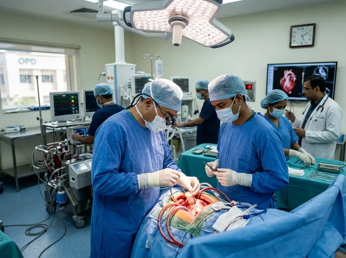
Aortic root disease often develops without overt clinical symptoms for many years. Consequently, clinicians frequently identify thoracic aortic dilation during incidental radiological scans. When the aortic root expands, surgical intervention aims to prevent rupture or acute dissection. For eligible individuals, the David procedure offers an alternative to composite graft replacement by preserving the native aortic valve. However, surgeons must evaluate patients before severe valvular deformation occurs. Early diagnosis creates a vital therapeutic window to protect cardiac function.
The Pathophysiology of Silent Root Dilatation
An expanding aneurysm insidiously alters the geometry of the aortic root. Specifically, progressive dilation stretches the sinotubular junction and the aortic annulus. In the early stages, the valve leaflets maintain adequate coaptation despite root enlargement. Therefore, patients usually remain completely asymptomatic and feel healthy.
However, unchecked annular dilation eventually induces significant aortic regurgitation. This chronic volume overload imposes severe diastolic wall stress on the left ventricle. Over time, progressive eccentric hypertrophy leads to irreversible myocardial dysfunction. Furthermore, delayed intervention reduces the likelihood of successful valve-sparing reconstruction. Once the leaflets stretch or prolapse excessively, surgeons must replace rather than repair the valve.
Preserving Function: Why the David Procedure Matters
Conventional management historically relied on the classic Bentall operation. In this composite replacement, the surgeon excises both the root and the native valve. Consequently, younger recipients receive mechanical prostheses requiring lifelong oral anticoagulation with warfarin. This necessity introduces persistent hazards of major haemorrhage and thromboembolism. Additionally, it significantly complicates pregnancy management in young women.
In contrast, the David procedure replaces the diseased aortic sinuses while resuspending the native valve within a vascular graft. This reimplantation technique restores physiologic aortic root geometry without prosthetic valve implantation. As a result, patients avoid long-term anticoagulation regimens and maintain superior haemodynamics. Moreover, preserved native valves demonstrate excellent durability across two decades when surgical teams achieve symmetric leaflet coaptation.
Patient Selection and Anatomical Criteria
Not every patient with root enlargement qualifies for reimplantation. Therefore, cardiologists must utilize detailed transthoracic and transoesophageal echocardiography during initial assessment. Surgeons evaluate leaflet pliability, calcification, symmetry, and effective height. In addition, contrast-enhanced computed tomography maps coronary anatomy and precise root dimensions.
Younger adults and patients with connective tissue disorders, such as Marfan syndrome, represent prime candidates. For these cohorts, early intervention avoids lifelong mechanical valve morbidity. Similarly, select individuals with non-calcified bicuspid aortic valves may undergo successful reconstruction. Conversely, surgeons typically recommend bioprosthetic replacement in patients over 70 years old. Ultimately, pristine leaflet anatomy determines eligibility rather than absolute aneurysm diameter alone.
Addressing Follow-Up Challenges in Indian Practice
Long-term surveillance remains essential to monitor valve durability and recurrent regurgitation. However, healthcare centres across India encounter substantial obstacles in tracking surgical cohorts. Many patients travel vast distances from rural communities for surgery and subsequently return home. Consequently, local contact details frequently change, which complicates active longitudinal tracking.
Furthermore, remote diagnostic facilities often produce non-standardized echocardiographic assessments. To address these gaps, tertiary centres now adopt structured electronic health records and telemedicine follow-up pathways. Digital platforms enable regional clinicians to share imaging directly with specialized aortic teams. Thus, continuous multidisciplinary coordination ensures early detection of leaflet deterioration and preserves long-term surgical success.
Frequently Asked Questions
Q1: What is the primary difference between the David procedure and the Bentall procedure?
The David procedure replaces the dilated aortic root while preserving the patient's native aortic valve leaflets. In contrast, the Bentall procedure replaces both the root and the valve with a prosthetic composite graft, requiring lifelong anticoagulation if a mechanical valve is used.
Q2: Why is early surgical referral critical for valve-sparing root replacement?
Early referral allows surgeons to operate before secondary annular stretching causes severe leaflet prolapse or irreversible ventricular dilation. Once leaflets suffer severe geometric damage, valve-sparing techniques become unfeasible, necessitating prosthetic replacement.
Q3: How does surveillance continue for rural patients treated in Indian tertiary centres?
Hospitals increasingly leverage telemedicine portals and centralized electronic registries. Local cardiologists capture echocardiographic images and transfer them to tertiary surgical teams, ensuring accurate serial monitoring without requiring arduous patient travel.
References
- Silent Aortic Disease Can Offer a Narrow Window to Save the Heart’s Own Valve - ETHealthworld
- David TE. Aortic valve sparing operations: outcomes at 20 years. Ann Cardiothorac Surg. 2013;2(1):24-29.
- Kelly JJ, Desai ND, Patrick WL, et al. Outcomes of aortic valve repair with the reimplantation technique and maintenance of commissural orientation. Ann Cardiothorac Surg. 2023;12(4):341-350.


