Novel MRI Dye Detects Tiny Lung Tumors Without Radiation
Novel MRI Dye Detects Tiny Lung Tumors Without Radiation
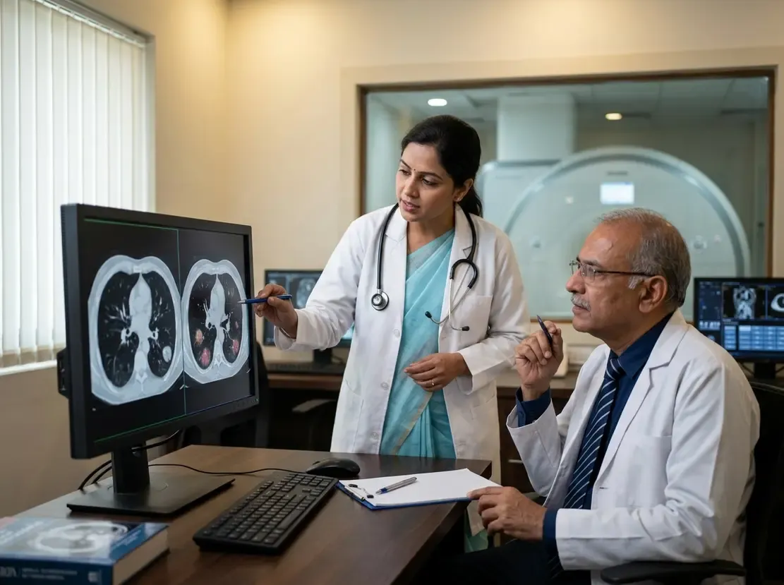
Early diagnosis remains the most formidable challenge in thoracic oncology today. Consequently, clinicians eagerly evaluate innovative diagnostic tools that identify microscopic primary lesions. A groundbreaking preclinical breakthrough in lung cancer MRI detection now promises to redefine early staging paradigms. Researchers engineered a novel protein-based contrast agent known as hProCA32.Collagen2 to visualize pulmonary nodules as small as 1 millimeter. Typically, standard diagnostic pathways discover lung cancer only after regional or distant dissemination occurs. Therefore, achieving sub-centimeter sensitivity without ionizing radiation could fundamentally transform screening algorithms. This innovative molecular imaging approach also evaluates intrinsic tumor biology, helping clinicians distinguish indolent lesions from highly invasive malignant phenotypes.
The Role of hProCA32.Collagen2 in Lung Cancer MRI Detection
The development of hProCA32.Collagen2 represents a substantial technical leap over conventional gadolinium-based agents. Specifically, the protein scaffold exhibits exceptionally high relaxivity at standard clinical magnetic field strengths. As a result, the agent provides robust image contrast even at minimal tissue concentrations. In preclinical animal models, researchers successfully detected both primary parenchymal lesions and hidden metastases across multiple visceral organs. In addition, the imaging revealed occult metastatic deposits within the adrenal glands and kidneys. Standard high-resolution computed tomography often misses these tiny anatomical alterations. Moreover, magnetic resonance imaging avoids repeated exposure to ionizing radiation, providing a safer longitudinal surveillance option for high-risk patients.
Targeting Extracellular Matrix Remodeling
Tumor progression relies heavily on structural alterations within the extracellular matrix. Specifically, malignant transformation stimulates excessive deposition and reorganization of type I collagen fibers around growing lesions. The experimental agent selectively targets this newly remodeled collagen scaffold. Consequently, the signal intensity directly reflects dynamic changes in the tumor microenvironment. This biological specificity enables clinicians to visualize aggressive cellular behaviors non-invasively. Furthermore, tumors bearing liver kinase B1 mutations demonstrate pronounced matrix remodeling and heightened invasive capacity. By highlighting these fibrotic margins, precision MRI provides vital prognostic insights before surgical resection. Thus, molecular imaging bridges the critical gap between macroscopic radiological detection and molecular pathology.
Clinical Implications for Oncology and Precision Staging
Integrating targeted MRI contrast into oncology practice may soon improve individualized management pathways. For instance, the technology could reduce the necessity for repeat invasive lung biopsies in frail individuals. Clinicians can accurately monitor therapeutic efficacy over time by assessing collagen structural shifts during chemotherapy or targeted interventions. However, translation from animal models to human clinical trials requires rigorous evaluation of safety, clearance kinetics, and tolerability. Biotech researchers currently prepare an investigational new drug application for regulatory review. If clinical trials confirm these preclinical findings, precision molecular MRI will expand screening options globally. Ultimately, early identification of aggressive metastases offers patients the best opportunity for curative therapeutic intervention.
Frequently Asked Questions
Q1: What is hProCA32.Collagen2 and how does it detect lung cancer?
It is an experimental protein-based MRI contrast agent that selectively binds to type I collagen. Aggressive lung tumors remodel this structural protein, enabling clear visualization on magnetic resonance imaging.
Q2: Why is lung cancer MRI detection advantageous over standard computed tomography?
Unlike conventional computed tomography, precision MRI eliminates exposure to ionizing radiation. Furthermore, the molecular agent can detect occult 1-millimeter metastases that standard computed tomography scans frequently miss.
Q3: When will this new contrast agent become available for clinical practice?
The agent is currently completing preclinical development, with researchers preparing an investigational new drug application. Consequently, human clinical trials must first evaluate safety, dosing, and imaging efficacy before regulatory approval.
References
- New dye for MRI exams detects lung cancer earlier - ETHealthworld
- Li D, Bamishaye O, Li S, et al. Early detection of invasive lung cancer and multiorgan metastasis by a collagen-targeted protein MRI contrast agent. Science Advances. 2026.
- European Medical Journal. Collagen Targeted MRI Detects Early Lung Cancer. EMJ Oncology. 2026.