Can Cervical Doppler Quantify Placenta Accreta Risk?
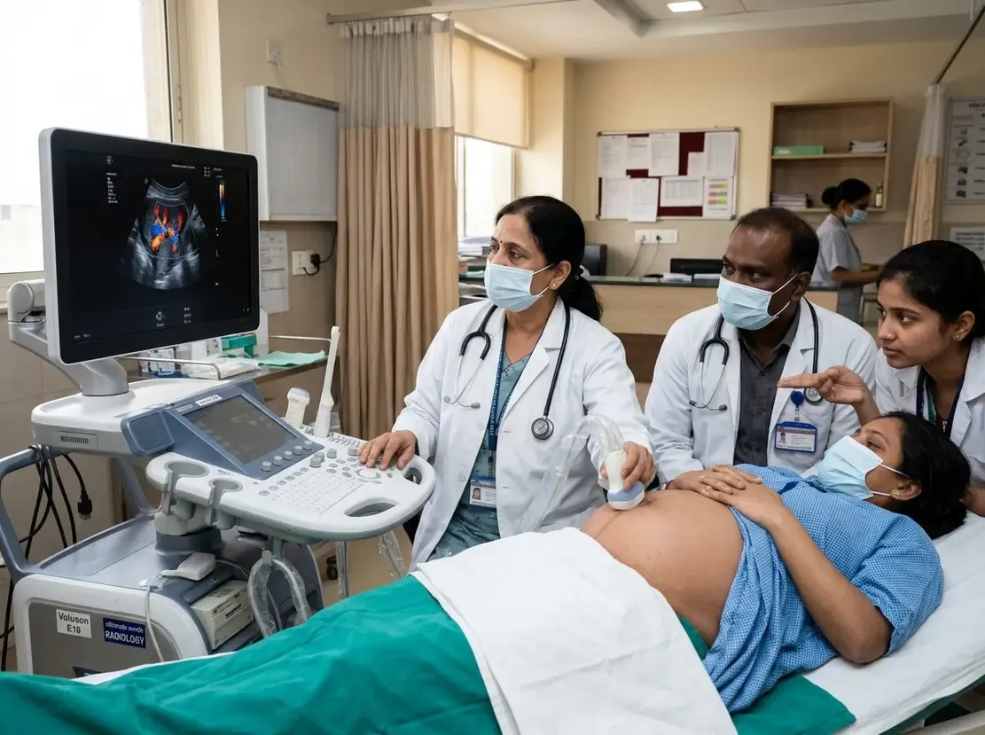
Diagnosing placenta accreta spectrum accurately remains a major clinical challenge in modern obstetrics. Early identification of abnormal invasive placentation allows multidisciplinary teams to plan complex surgeries and minimize catastrophic maternal bleeding. Therefore, objective imaging biomarkers are vital for improving clinical decision-making before delivery.
Assessing Placenta Accreta Spectrum with Quantitative Ultrasound
Traditional ultrasound evaluation relies primarily on subjective sonographic markers. These markers include placental lacunae, loss of the clear zone, and abnormal hypervascularity. However, subjective assessment introduces considerable interobserver variation. Consequently, researchers at the University of Texas evaluated a standardized quantitative vascularization method. Specifically, the team examined the anterior fornix and cervix region using transvaginal color Doppler imaging. They analyzed videoclips through post-processing software to calculate the mean percentage of color pixels across sequential frames.
Study Findings and Diagnostic Accuracy
The prospective study evaluated 117 pregnant women presenting with anterior placenta previa at elevated risk for morbid adherence. Histopathology subsequently confirmed placenta accreta spectrum in 89 patients. Notably, the quantitative vascularization technique demonstrated an area under the curve of 0.821 for discriminating invasive cases. Furthermore, investigators identified an optimal cutoff value of 8.5%. This threshold yielded a sensitivity of 70.5% and a specificity of 89.7%. In addition, the positive predictive value reached 93.9%, demonstrating exceptional diagnostic reliability.
Surgical Outcomes and Blood Loss Prediction
Severe intraoperative hemorrhage represents the most perilous complication of invasive placentation. In this prospective cohort, 53 patients experienced intraoperative blood loss exceeding 1500 mL. Importantly, quantitative vascularization values of 8.5% or greater correlated with significantly increased odds of severe hemorrhage. Patients exceeding this threshold also required greater volumes of transfused blood products. Thus, this non-invasive metric provides crucial risk stratification before surgical intervention.
Frequently Asked Questions
Q1: What is quantitative vascularization of the cervix?
It is an ultrasound technique that calculates the percentage of color Doppler flow pixels within the anterior fornix and cervix region.
Q2: How does this ultrasound metric improve patient management?
The metric objectively predicts placental invasion severity and anticipated surgical hemorrhage, enabling timely surgical planning and blood product preparation.
References
- Kassir E et al. Association of quantitative vascularization of the anterior fornix/cervix region with placenta accreta spectrum and associated intraoperative hemorrhage. Ultrasound Obstet Gynecol. 2026 Oct 25. doi: 10.1002/uog.70362. PMID: 42927468.
- Jauniaux E, Bhide A, Kennedy A, Woodward P, Hubinont C, Collins S; FIGO Placenta Accreta Diagnosis and Management Expert Consensus Panel. FIGO consensus guidelines on placenta accreta spectrum disorders: Prenatal diagnosis and screening. Int J Gynaecol Obstet. 2018;140(3):274-280.