
AIIMS Defines Ray's Triangle in Complex Cancer Surgery
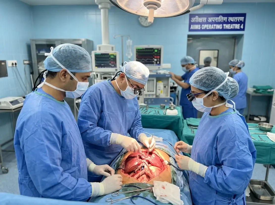
Surgical oncologists at AIIMS New Delhi have identified Ray's triangle as a critical anatomical warning zone during upper abdominal cancer operations. Specifically, this high-risk area sits at the confluence of the liver, the inferior vena cava, and the right hepatic vein. Dr M D Ray and Dr Anik Rathee described this landmark in the Journal of the Egyptian National Cancer Institute. Consequently, recognizing this anatomical landmark helps surgeons clear occult metastatic disease without causing catastrophic vascular injuries.
The Surgical Significance of Ray's Triangle
Complete cytoreduction offers the strongest predictor of overall survival in advanced ovarian carcinoma. However, malignant cells frequently lodge in occult upper abdominal spaces, especially behind the liver and along the diaphragm. Surgeons must completely mobilise the liver to inspect and resect these hidden tumor deposits. Therefore, extensive mobilization remains essential to achieve complete macroscopic tumor clearance. In this AIIMS cohort of 205 ovarian cancer patients, surgical teams achieved complete or near-complete cytoreduction in 94.6% of cases. In fact, surgeons accomplished complete tumor resection in 178 patients, while only 16 retained minimal residual disease.
Mitigating Hepatic Vein Injury and Vascular Hazards
However, right upper quadrant cytoreduction carries significant vascular hazards during extensive mobilization. Notably, researchers observed right hepatic vein lacerations in nine patients, representing an overall injury rate of 4.4%. These venous injuries occurred predominantly during the initial phase, affecting seven of the first 100 cases. In contrast, the injury incidence dropped sharply to 1.9% across the subsequent 105 patients. Patients who experienced vascular tears suffered greater operative hemorrhage and required extended intensive care stays. Furthermore, dense post-chemotherapy adhesions led surgeons to abort dissection in two patients to prevent severe bleeding.
Best Practices for Safe Upper Abdominal Dissection
The authors emphasize that Ray's triangle represents a practical surgical warning zone rather than a formal anatomical entity. Accordingly, surgical teams must follow a disciplined, systematic technique rather than relying on forceful or blind tissue dissection. Surgeons initiate exposure through a midline laparotomy and progressively divide hepatic ligaments to visualize retrohepatic vascular structures. Additionally, teams must identify retrohepatic vessels early before applying traction to the mobilized liver. Ultimately, structured dissection protocols protect crucial venous anatomy while maximizing the likelihood of complete macroscopic tumor clearance.
Frequently Asked Questions
Q1: What is Ray's triangle in abdominal cancer surgery?
Ray's triangle is a surgical warning area where the liver, the inferior vena cava, and hepatic vein meet. AIIMS oncologists identified this zone as a high-risk site for vascular injury during extensive liver mobilization.
Q2: Why is complete liver mobilization necessary in advanced ovarian cancer?
Malignant cells frequently lodge in occult spaces behind the liver and along the right diaphragm. Therefore, surgeons mobilise the liver to detect and excise these hidden deposits, ensuring complete cytoreduction.
Q3: How can surgeons avoid major venous injuries around this landmark?
Consequently, surgeons prevent injuries by employing systematic sharp dissection, avoiding blind traction, and identifying venous structures early.
References
- AIIMS experts identify 'Ray's triangle' as high-risk area in complex abdominalcancer surgery - ETHealthworld
- Ray MD, Rathee A. Performing right upper abdominal cytoreduction: anatomical planes, liver mobilization and occult spaces. Journal of the Egyptian National Cancer Institute. 2026.
- Shin W, Park SY, Lim MC, et al. Narrative review of liver mobilization, diaphragm peritonectomy, full-thickness diaphragm resection, and reconstruction. Gland Surgery. 2021;10(3):1212-1224.


