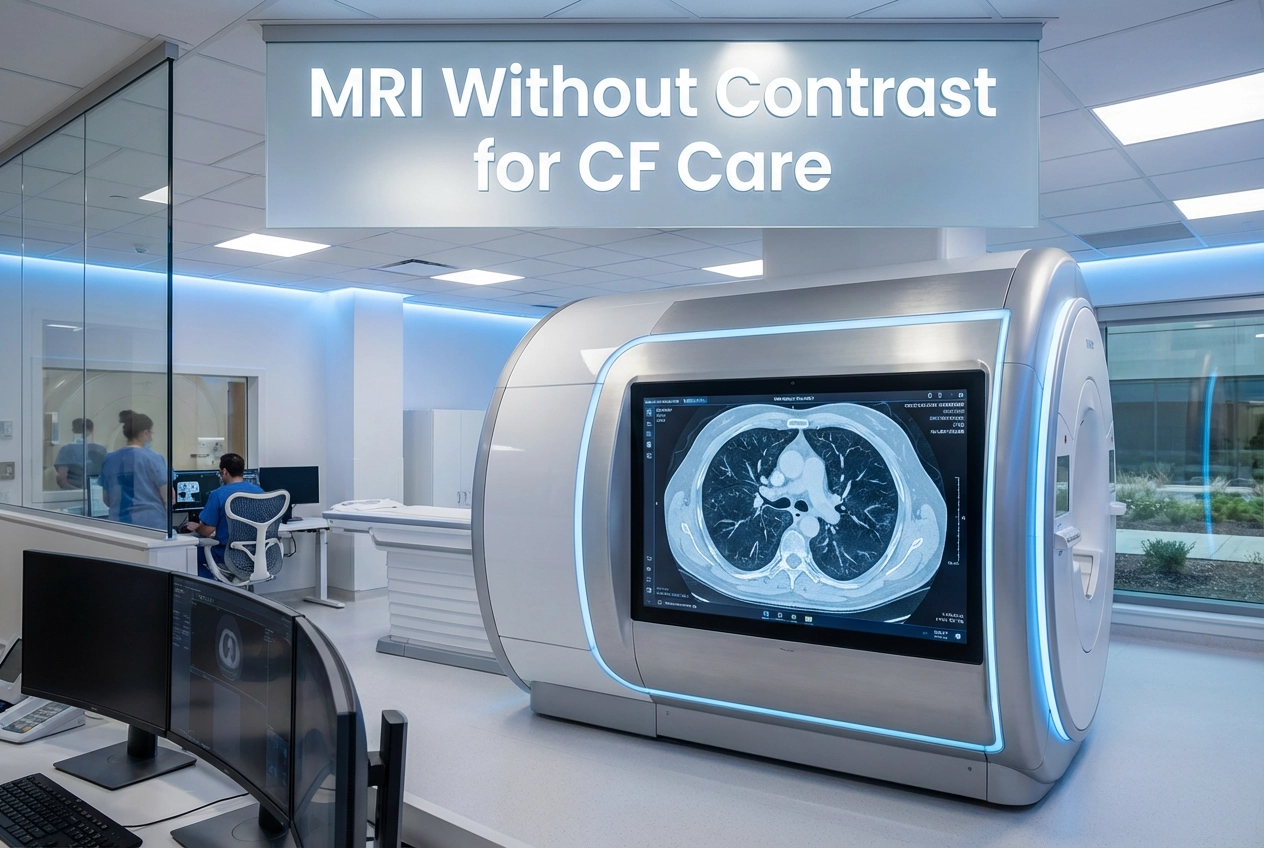
The ability to accurately monitor lung disease progression is essential for people with cystic fibrosis (pwCF). In this context, CF Pleural MRI has become a vital, radiation-free tool for longitudinal surveillance. However, the use of intravenous (IV) contrast media has remained standard practice for assessing inflammation and thickening of the pleura. This standard posed a challenge, as IV contrast introduces costs, preparation time, and potential risks, including allergic reactions and gadolinium deposition. Therefore, new research sought to determine if simpler, unenhanced MRI sequences could provide equivalent diagnostic information.
Unenhanced Sequences Match Diagnostic Value in CF Pleural MRI
A new multi-reader study, which included 75 pwCF, directly compared different unenhanced and contrast-enhanced sequences for pleural evaluation. Researchers used the validated morpho-functional chest MRI score for this assessment. Initially, contrast-enhanced T1-weighted sequences with fat saturation (T1CEFS) served as the diagnostic benchmark. The results revealed a strong correlation between the standard contrast-enhanced sequence and unenhanced T1-weighted sequences. Specifically, the unenhanced, fat-saturated T1-weighted sequence (T1UEFS) showed the highest agreement with the T1CEFS regarding the total pleural findings score. Thus, the diagnostic value of the contrast-agent-free sequence was effectively equivalent to the gold standard. Interestingly, the study noted that a standard T2-weighted sequence tended to overestimate findings, while a plain unenhanced T1-weighted sequence tended to underestimate them.
Clinical Impact: Streamlining Care and Enhancing Safety
The validation of the contrast-free T1UEFS sequence is the most significant takeaway for clinical practice. Consequently, doctors can now assess CF-related pleural inflammation and thickening just as precisely without administering an IV contrast agent. Furthermore, eliminating the need for contrast media streamlines the imaging protocol significantly. Besides simplifying the process, this change reduces the patient burden associated with IV access and waiting periods after injection. More importantly, it removes the risks associated with gadolinium-based contrast agents (GBCAs). These risks include allergic reactions or the potential for nephrogenic systemic fibrosis (NSF) in patients with underlying kidney concerns. Ultimately, this finding paves the way for future routine MRI monitoring for pwCF that consistently avoids contrast agents, especially for long-term follow-up and pediatric patients.
Frequently Asked Questions
Q1: What is the main finding of the study regarding CF Pleural MRI?
Unenhanced, fat-saturated T1-weighted sequences (T1UEFS) provide diagnostic value equivalent to contrast-enhanced T1-weighted sequences for assessing pleural inflammation and thickening in people with cystic fibrosis (pwCF).
Q2: Why is eliminating intravenous contrast media beneficial for pwCF?
The elimination of IV contrast media streamlines the MRI monitoring process, reduces patient burden by avoiding IV access and post-injection waiting times, and removes the risk of gadolinium-related complications like allergic reactions or nephrogenic systemic fibrosis (NSF).
References
- Schobert IT et al. MRI scoring of pleural findings in cystic fibrosis does not require intravenous contrast media. Eur Radiol. 2025 Dec 23. doi: 10.1007/s00330-025-12209-5. PMID: 41436642.
- Dinh, H. R., & Szejnfeld, D. (2025). Safety issues related to intravenous contrast agent use in magnetic resonance imaging. ResearchGate.
- Thiele H, Puderbach M, Biederer J. Chest magnetic resonance imaging in cystic fibrosis: technique and clinical benefits. J Thorac Dis. 2022 Nov;14(11):4498-4508.