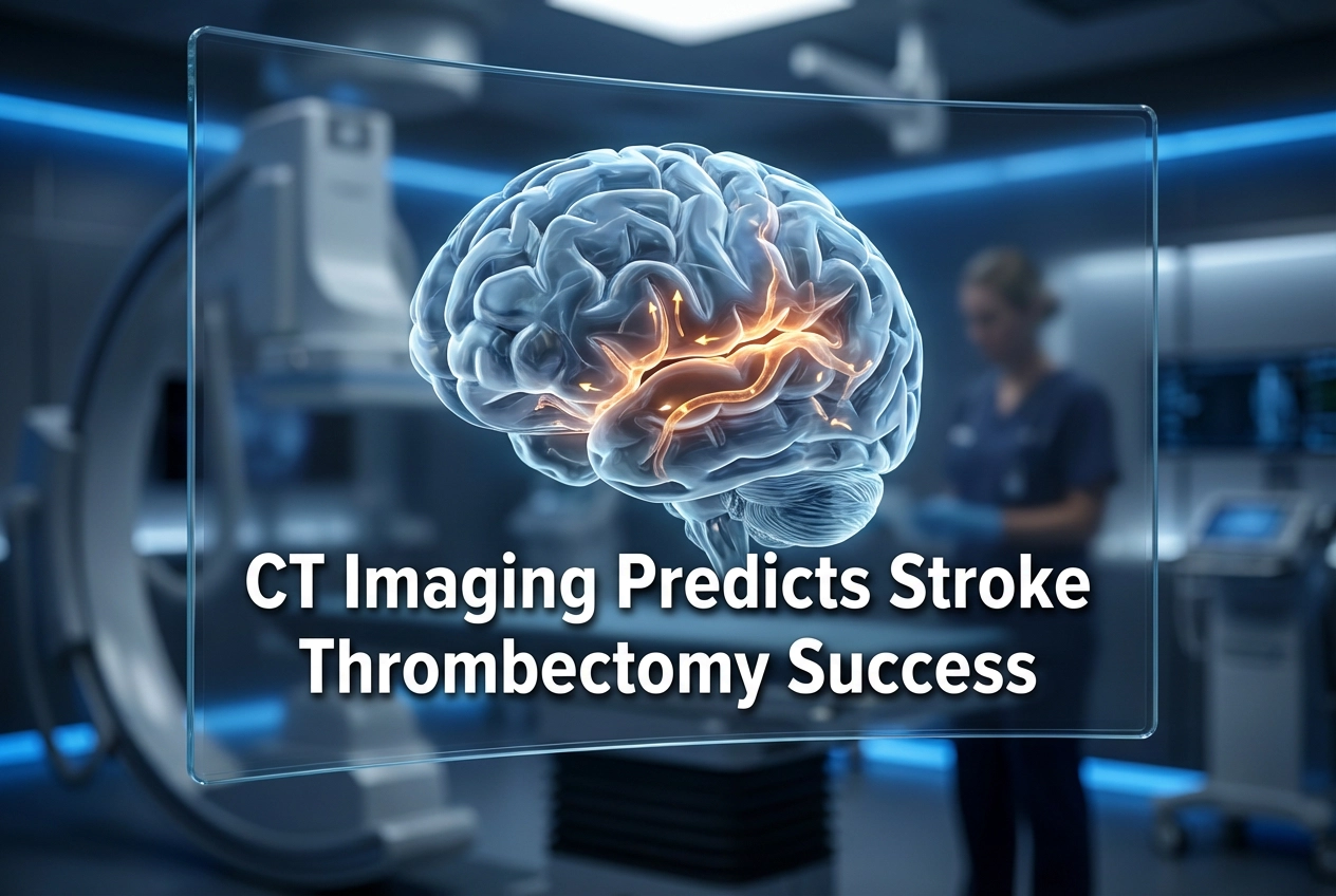
Endovascular thrombectomy (EVT) represents the standard of care for acute ischemic stroke caused by anterior large vessel occlusion (LVO). However, accurately predicting which patients will experience a favorable functional outcome (mRS 0–2) remains a critical challenge. For this reason, new research investigated the value of baseline CT imaging for EVT prediction stroke outcome, finding that imaging data holds considerable predictive power alone.
CT Imaging’s Role in EVT Prediction Stroke Outcome
The study utilised individual patient data from seven randomised EVT trials, analysing 1,391 patients with anterior LVO. The median age of participants was 67 years. Researchers designed a prediction model focusing solely on baseline stroke-related and brain frailty CT imaging features. Subsequently, the model’s performance for predicting both a good functional outcome and the benefit of EVT was assessed.
Notably, the CT imaging-only model demonstrated substantial discrimination for predicting a good functional outcome, registering a C-statistic of 0.700. Additionally, its discrimination for treatment benefit achieved a C-for-benefit of 0.640. These findings strongly suggest that early CT imaging provides vital information about a patient’s prognosis, even without advanced clinical data.
Optimising Prediction: Imaging Plus Clinical Data
Although CT imaging alone proved valuable, the study aimed to create the most robust model possible. Therefore, scientists incorporated the strongest clinical predictors: patient age and the National Institutes of Health Stroke Scale (NIHSS) score. Adding these limited clinical characteristics significantly improved the model’s overall predictive performance. The C-statistic for a good functional outcome increased to 0.733. Likewise, the C-for-benefit for predicting treatment advantage rose to 0.675.
In comparison to the established MR PREDICTS model, the combined clinical-imaging model achieved a predictive performance that was only slightly lower. Therefore, a simple combination of readily available non-contrast CT features and basic clinical scores can nearly match the performance of more complex predictive tools. This approach offers practical benefits for rapid, real-world clinical decision-making across stroke centres.
The Clinical Relevance for LVO Stroke Management
For stroke physicians, this research confirms that baseline CT imaging should be the central component of initial patient assessment. Furthermore, it supports the current practice of incorporating clinical variables like age and NIHSS in the triage process. Recognising the potential of CT, other models are also exploring its role. For example, some AI algorithms now use CT angiography (CTA) to rapidly detect LVOs, aiming to decrease time-to-treatment. Furthermore, quantitative CT measures, such as Net Water Uptake (NWU), may help determine which patients with large ischemic lesions will benefit from EVT, a crucial factor in extended time window treatment. Consequently, evolving CT-based biomarkers and algorithms continue to refine patient selection for this life-saving procedure.
Frequently Asked Questions
Q1: What specific CT features were used to predict EVT outcome in the study?
The prediction model included baseline stroke-related features and brain frailty features derived from the CT imaging. These are typically indicators of established infarct size (e.g., ASPECTS score) and baseline brain health.
Q2: How did the study define a “good functional outcome” for stroke patients?
The researchers defined a good functional outcome as a modified Rankin Scale (mRS) score of 0 to 2. This range signifies functional independence 90 days after the stroke event.
Q3: Why is combining clinical and imaging data better than using CT alone?
While CT imaging provides essential information about the brain’s physical state, clinical data like age and stroke severity (NIHSS score) account for the patient’s overall health and physiological resilience. Combining both offers a more complete picture, resulting in the best predictive performance for a patient’s response to endovascular thrombectomy.
References
- Luijten SP et al. A CT imaging-based prediction model of functional outcome and benefit of endovascular thrombectomy for ischemic stroke. Eur Radiol. 2026 Jan 14. doi: 10.1007/s00330-025-12207-7. PMID: 41533062.
- Broocks G et al. Degree of net water uptake on CT helps predict EVT treatment efficacy. Radiology. 2025 Nov 4.
- Stib et al. Detecting Large Vessel Occlusion at Multiphase CT Angiography by Using a Deep Convolutional Neural Network. Radiology. 2020 Sep 29.