The diagnosis of Fetal Bradycardia LQTS presents a significant challenge in prenatal cardiology. Consequently, a new study offers crucial insights into the association between specific fetal heart rhythms and congenital Long QT Syndrome (LQTS). The research focused on fetuses presenting with isolated sinus bradycardia, 2:1 atrioventricular block (AVB), or ventricular tachycardia (VT). They excluded cases involving major congenital heart disease or maternal autoimmune antibodies (anti-Ro/La). Therefore, this research isolates the cardiac channelopathy component.
Understanding Fetal Bradycardia LQTS Presentation
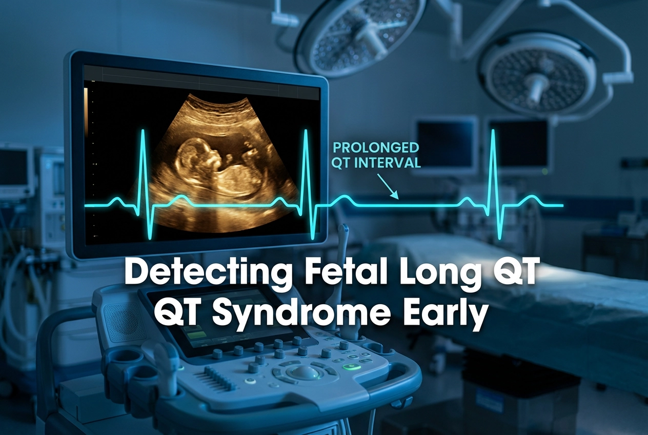
Fetal bradycardia is defined as a ventricular rate more than 2 SD below the mean for the corresponding gestational age. Moreover, this study recognized three primary rhythm presentations potentially linked to fetal LQTS: sinus bradycardia, functional 2:1 AVB, and polymorphic VT. The study reviewed 22 fetuses referred to a tertiary fetal cardiology center. Interestingly, none of the included cases had a prior family history of LQTS. For instance, the median presenting fetal heart rate was 120 bpm, corresponding to a Z-score of -3.04 in sinus rhythm.
The investigators used the left ventricular isovolumetric relaxation time (LVIRT) and its normalized proportion (N-LVIRT) to estimate the QT interval. In fact, these measures are considered important diagnostic tools for LQTS in utero. However, N-LVIRT and LVIRT were above the threshold values in only a minority of the genetically confirmed cases (two and six cases, respectively). Therefore, direct genetic confirmation remains paramount for diagnosis.
The Critical Role of Prenatal Genetic Testing
The key finding reveals that LQTS is the most common underlying cause for persistent sinus bradycardia, provided major congenital heart disease and autoimmune disease are absent. Consequently, clinicians must maintain a high index of suspicion for this diagnosis, which often involves a multidisciplinary team approach. Genetic testing was performed in 14 cases, 12 of which received testing prenatally. This testing identified a positive LQTS genotype in 13 cases. The most frequent genotypes were KCNQ1 (eight cases), which is a common finding in LQTS cohorts, followed by KCNH2 (two cases). KCNE1, SCN5A, and CALM2 each accounted for one case.
Furthermore, prenatal genetic confirmation enables a tailored approach to parental counseling. This early diagnosis allows for specialized management planning both during the pregnancy and immediately postpartum. Ultimately, this precision medicine approach can significantly improve the clinical outcome for the neonate. In India, molecular analysis of these common LQTS genes (KCNQ1, KCNH2, SCN5A) is well-established, validating the utility of this diagnostic strategy.
Frequently Asked Questions
Q1: What is the clinical definition of fetal bradycardia used in this study?
Fetal bradycardia is defined as a ventricular rate that falls more than two standard deviations below the gestational-age-specific mean.
Q2: What is the most common underlying diagnosis for persistent fetal sinus bradycardia without major congenital heart disease?
The most common finding in fetuses presenting with persistent sinus bradycardia, in the absence of major congenital heart disease or autoimmune disease, is Long QT Syndrome (LQTS).
Q3: Why is prenatal genetic testing important in cases of suspected Fetal Bradycardia LQTS?
Prenatal genetic testing enables clinicians to provide tailored parental counseling and allows for specialized management planning of the pregnancy and delivery, leading to better neonatal outcomes.
References
- Chivers S et al. Prenatal presentation of fetal bradycardia and long QT syndrome. Ultrasound Obstet Gynecol. 2026 Jan 03. doi: 10.1002/uog.70132. PMID: 41482982.
- Stramba-Badiale M et al. The natural history of fetal long QT syndrome. J Am Heart Assoc. 2022. doi: 10.1161/JAHA.122.025805. PMID: 36474163.
- Rajamani K et al. Phenotype guided characterization and molecular analysis of Indian patients with long QT syndromes. J Cardiovasc Electrophysiol. 2017. doi: 10.1111/jce.13110. PMID: 27928875.