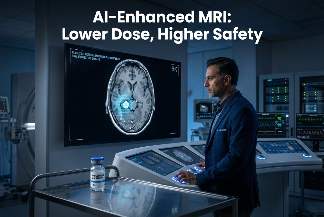
The critical need for gadolinium dose reduction in contrast-enhanced MRI (CE-MRI) is driving significant innovation. A new deep learning (DL) model demonstrates the potential to dramatically lower the required contrast agent dose for imaging the cerebellopontine angle (CPA) cistern. This advancement maintains diagnostic quality for conditions like vestibular schwannoma (VS). Experts acknowledge that this technology directly addresses the growing concern of gadolinium retention in the body. Consequently, this deep learning approach provides a valuable path to enhanced patient safety during neuroimaging.
Deep Learning Restores Low-Dose Contrast-Enhanced MRI
Researchers retrospectively analyzed 203 MRI studies from 72 VS patients. They simulated low-dose CE-MRI scans using various reductions of the contrast agent. Therefore, DL models were trained to restore these simulated low-dose images back to a standard-dose quality. The goal was specifically to evaluate the diagnostic utility and image quality of the DL-restored T1ce images. Both image quality metrics, such as structural similarity index and peak signal-to-noise ratio, and clinical performance, like segmentation accuracy, were evaluated. The DL-restored T1ce from a 10% input dose showed excellent image quality. Furthermore, the 30% input dose restoration was rated even more informative by a head and neck radiologist. The diagnostic characterization remained possible even with only 10–30% of the standard contrast dose.
Accuracy and Clinical Relevance of Gadolinium Dose Reduction
The DL model significantly improved the quality of the low-dose images. For instance, at a 10% input dose, the segmentation accuracy for VS improved markedly. Specifically, the Dice coefficient increased from 0.673 to 0.734. The 95% Hausdorff distance also decreased from 2.38 mm to 2.07 mm. Similarly, the average surface distance improved from 1.00 mm to 0.59 mm. These metrics confirm the model’s ability to create diagnostically sufficient images from minimal contrast data. The study validates that low-dose MRI of the CPA cistern, when processed with DL, enables accurate lesion detection and diagnostic characterization. Consequently, this technology can substantially reduce the use of gadolinium-based contrast agents (GBCAs). This is clinically important because GBCAs, while generally safe, are linked to gadolinium retention in the brain and other tissues.
Global Impact of Reduced Gadolinium MRI on Patient Safety
Globally, healthcare providers have focused on strategies to minimize GBCA exposure due to concerns over gadolinium retention and Nephrogenic Systemic Fibrosis (NSF) risk in vulnerable patients. Therefore, the development of DL-based techniques represents a major step forward. Other studies support this finding, demonstrating that AI can reduce the gadolinium dose 10-fold for general brain MRI while maintaining diagnostic value. Thus, this new technique for CPA cistern imaging aligns with a broader trend in neuroradiology. The use of this DL model provides an excellent opportunity to enhance patient safety. Furthermore, it helps clinicians make informed decisions about complex cases like vestibular schwannoma diagnosis and surveillance. This methodology ensures diagnostic clarity while proactively mitigating potential long-term risks associated with gadolinium administration, especially in patients requiring multiple follow-up scans.
Frequently Asked Questions
Q1: Why is gadolinium dose reduction important?
It is important because it mitigates potential risks associated with gadolinium-based contrast agents (GBCAs), such as gadolinium retention in the body, while still providing the necessary image clarity for diagnosis.
Q2: What percentage of the standard contrast dose is needed with the deep learning model?
The study found that the deep learning model can restore diagnostic quality images from low-dose scans using only 10% to 30% of the standard contrast agent dose.
Q3: Which conditions benefit from this deep learning MRI approach?
This specific study focused on vestibular schwannoma patients, showing that the DL-restored images of the cerebellopontine angle cistern were sufficient for diagnosis and management.
References
- Chen Y et al. A deep learning model to reduce agent dose for contrast-enhanced MRI of the cerebellopontine angle cistern. Eur Radiol. 2025 Dec 02. doi: 10.1007/s00330-025-12187-8. PMID: 41329327.
- Gong E et al. Deep learning enables reduced gadolinium dose for contrast-enhanced brain MRI. J Magn Reson Imaging. 2018;48:330–340.
- Gadolinium Retention: A Research Roadmap from the 2018 NIH/ACR/RSNA Workshop on Gadolinium Chelates. Invest Radiol. 2018;53(4):185-195.