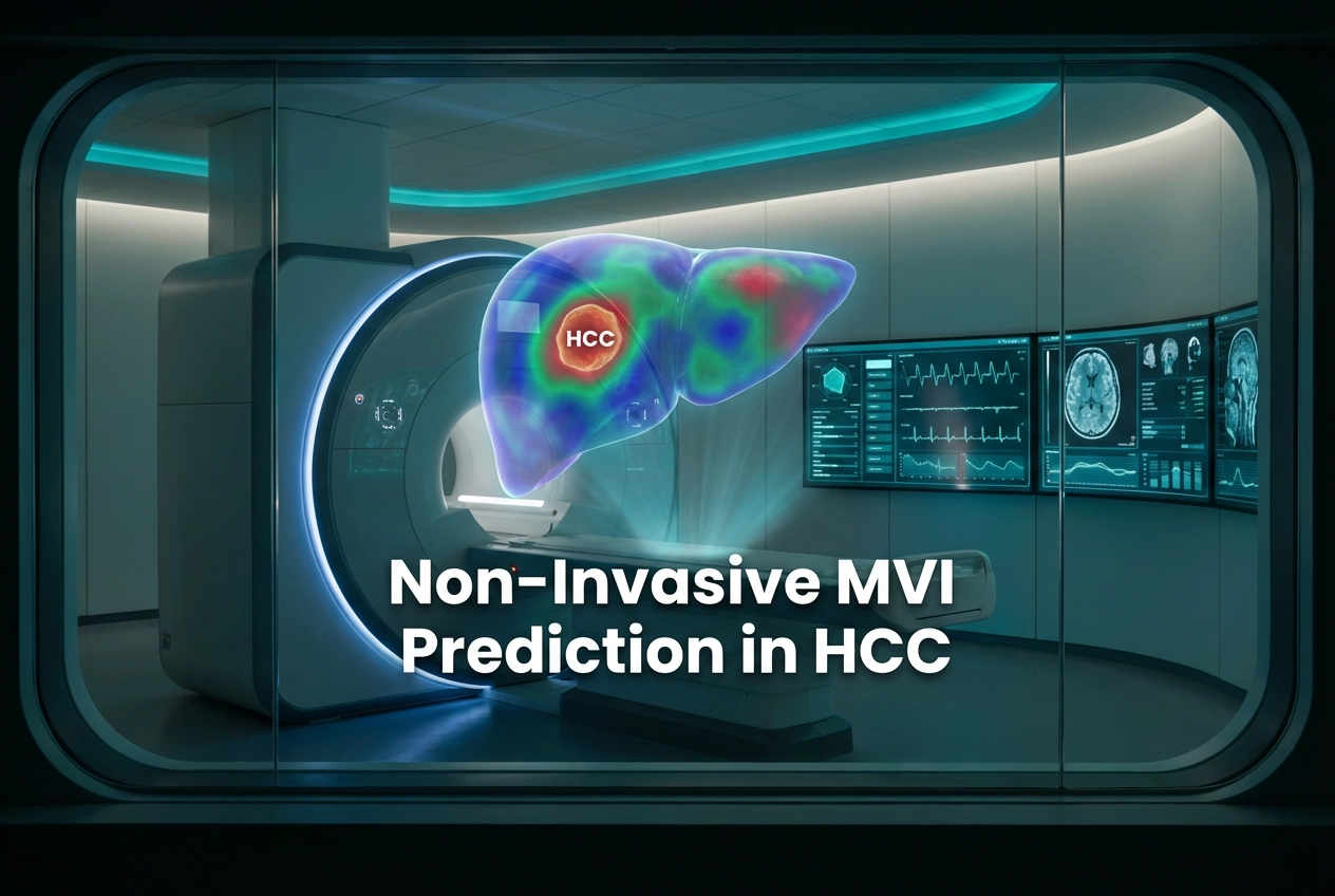
Microvascular invasion (MVI) is a strong, independent prognostic factor in patients diagnosed with hepatocellular carcinoma (HCC). Furthermore, the accurate, preoperative assessment of MVI is critical for determining the most effective treatment strategy, such as surgical resection versus liver transplantation. This study specifically explores the diagnostic value of mechanical parameters measured by compression MR elastography (MRE) for HCC MVI Prediction.
Understanding MR Elastography’s Role in HCC MVI Prediction
Preoperative imaging plays a vital role in identifying high-risk HCC patients. Conventional imaging methods often struggle to non-invasively detect MVI. However, MRE, which assesses tissue stiffness and viscoelastic properties, offers a new approach. Consequently, this study investigated MRE parameters, including the tumor stiffening slope and basal visco-elastic parameters (storage modulus, loss modulus, and phase angle). These measurements were taken during both expiration and inspiration, capturing the effects of respiratory-induced compression.
Key Findings on Stiffening and Visco-Elasticity
The final study group included 53 patients with complete data, 31 of whom were confirmed to have MVI through histopathological examination after surgical resection. Specifically, the results showed a significant difference in certain MRE parameters between tumors with and without MVI (p < 0.05). Tumors without MVI exhibited a significantly higher compression stiffening slope. The storage modulus difference between inspiration and expiration also proved to be significantly higher in patients without MVI. Therefore, the compression stiffening slope emerged as the most effective single parameter for predicting MVI, achieving an impressive area under the curve (AUC) of 0.77. However, the combination of the stiffening slope and the basal storage modulus further improved diagnostic accuracy. This composite model yielded an AUC of 0.81, indicating excellent performance.
Clinical Implications for Surgical Planning
The ability to non-invasively and accurately predict MVI preoperatively holds significant clinical value. MVI detection strongly influences the risk of tumor recurrence and overall survival. Consequently, knowing the MVI status helps surgeons tailor the extent of resection and decide on the appropriateness of liver transplantation. Furthermore, this technique offers a promising, non-invasive means to stratify HCC patients before surgery. Therefore, it may help guide personalized treatment decisions for those at high risk for recurrence.
Frequently Asked Questions
Q1: Why is Microvascular Invasion (MVI) important in HCC?
MVI is considered a strong prognostic factor in hepatocellular carcinoma because its presence significantly increases the risk of tumor recurrence and shortens overall patient survival following treatments like surgical resection.
Q2: Which MR elastography parameter was most predictive of MVI?
The compression stiffening slope, which reflects non-linear elasticity during respiratory-induced compression, was the most effective single parameter for predicting MVI, showing an area under the curve (AUC) of 0.77.
References
- Pagé G et al. MR elastography in patients with hepatocellular carcinoma: tumor stiffening during compression induced by respiration to assess microvascular invasion. Eur Radiol. 2026 Feb 01. doi: 10.1007/s00330-025-12164-1. PMID: 41621038.
- HCC Management in India: Prognostic Factors and Treatment Strategies.
- Emerging Non-invasive Techniques for Microvascular Invasion Prediction in Hepatocellular Carcinoma.