Why Radiologists Miss Lung Nodules and How to Fix It
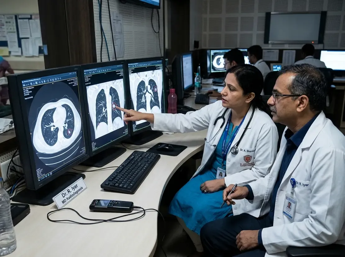
Diagnostic errors in thoracic imaging represent a pressing challenge in modern medicine. In particular, missed pulmonary nodules on chest CT scans remain a primary cause of malpractice claims and delayed diagnoses worldwide. Identifying these subtle lesions early can save lives, especially in high-burden clinical settings. Consequently, radiologists and clinicians must understand the mechanisms that drive these perceptual and diagnostic oversights.
Anatomic and Morphologic Determinants of Missed Pulmonary Nodules
Nodule characteristics strongly influence whether a radiologist detects an abnormality. For example, small lesions measuring under six millimeters frequently escape notice during routine review. Furthermore, pure ground-glass nodules present subtle attenuation differences that blend into normal parenchyma. Studies also show that surrounding anatomy complicates visual detection. Specifically, nodules nestled within endobronchial or perivascular spaces create confusing borders. Similarly, apical and subpleural regions conceal lesions behind dense vascular structures or ribs. Therefore, complex anatomical backgrounds significantly reduce lesion conspicuity.
Technical Limitations and Reader Fatigue
Beyond morphology, technical scanning parameters play a critical role. For instance, thick CT slices obscure tiny lesions through partial volume averaging. In addition, low-dose protocols and respiratory motion artifacts often degrade image clarity. Nevertheless, human cognitive limitations represent an even larger contributor to diagnostic error. Radiologists often suffer from visual fatigue during long reading shifts. Consequently, tired readers experience reduced visual search efficiency and inattentional blindness. Moreover, satisfaction of search can cause a clinician to stop reviewing after finding an initial abnormality. Thus, heavy workloads amplify cognitive blind spots during routine interpretations.
The Role of Artificial Intelligence and Medicolegal Implications
Modern artificial intelligence tools offer promising support for chest imaging. In fact, deep learning algorithms provide an effective second pair of eyes that highlights occult nodules. However, automated systems cannot replace careful human judgment. Some algorithms generate false positives that increase reader workload. Furthermore, current legal systems judge diagnostic errors against vague concepts of obviousness. Retrospective reviews often impose hindsight bias on clinicians. Therefore, the medical community requires objective benchmarks to define realistic detection standards. Adopting systematic scanning protocols helps radiologists defend their findings while improving thoracic patient care.
Frequently Asked Questions
Q1: Why are pure ground-glass nodules frequently overlooked on CT?
Pure ground-glass nodules have faint, hazy attenuation that easily blends with surrounding lung parenchyma. In addition, their subtle contrast makes them far less noticeable than dense solid lesions.
Q2: How does satisfaction of search contribute to radiologic errors?
Satisfaction of search occurs when a physician identifies one notable abnormality and prematurely halts further visual inspection. Consequently, the reader misses secondary findings, including subtle early-stage pulmonary nodules.
Q3: Can artificial intelligence completely eliminate missed nodules on chest CT?
Although artificial intelligence enhances detection sensitivity, it cannot eliminate errors completely. Radiologists must still verify all algorithm outputs to filter out false positives and evaluate anatomical context accurately. For further skill enhancement in this field, professionals often pursue advanced radiology training programs.
References
- Waite S et al. Missed Pulmonary Nodules on CT: Determinants and Implications for Radiologic Practice. AJR Am J Roentgenol. 2026 Nov 10. doi: 10.2214/AJR.26.35355. PMID: 42584409.
- Drew T, Võ ML, Wolfe JM. The invisible gorilla strikes again: Sustained inattentional blindness in expert observers. Psychol Sci. 2013;24(9):1848-1853.
- Hossain R, Wu CC, de Groot PM, Carter BW, Gilman MD, Abbott GF. Missed lung cancer. Radiol Clin North Am. 2018;56(3):365-375.