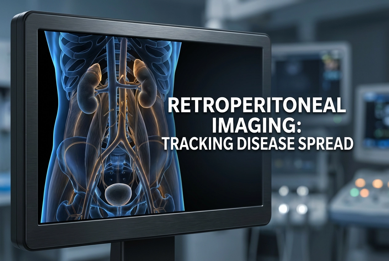
The retroperitoneum and pelvic extraperitoneum are intricate anatomic regions. These spaces contain numerous vital structures, for example, major vessels, lymphatics, nerves, and abdominopelvic organs. Consequently, these regions are highly susceptible to diverse pathologic processes. Retroperitoneal imaging, specifically CT and MRI, is indispensable for characterizing pathology and guiding management. Furthermore, the retroperitoneum represents the abdominal extraperitoneum, extending from the diaphragm superiorly to the pelvic brim inferiorly. Understanding the anatomy, imaging features, and patterns of disease spread within these spaces is critical for accurate diagnosis and effective disease management.
Retroperitoneal Imaging: Compartments and Anatomy
Fascial planes divide the extraperitoneal spaces into distinct compartments with defined contents and boundaries. These divisions effectively guide the spread of disease within and beyond the abdominopelvic cavity. The anterior and posterior renal fasciae (Gerota’s and Zuckerkandl’s fascia, respectively) form three major divisions in the retroperitoneum. Therefore, we recognize the anterior pararenal space, the perirenal space, and the posterior pararenal space. Pathologic fluid, for example, can track along these fascial planes. Moreover, infectious and inflammatory processes often follow these boundaries closely. The pelvic extraperitoneum, conversely, is located below the peritoneal reflection. It surrounds the pelvic organs and is also known as the subperitoneal space. Understanding these fascial barriers is critical for accurate radiologic diagnosis.
Pathways of Extraperitoneal Disease Spread
The retroperitoneum and pelvic extraperitoneum are vulnerable to various pathologic conditions, including congenital, inflammatory, infectious, neoplastic, and traumatic diseases. Imaging, such as CT and MRI, is fundamental for characterizing the nature of these processes. Moreover, disease spread in these compartments follows predictable patterns. Malignancies, for instance, typically disseminate via lymphatic and hematogenous routes. Inflammatory and infectious processes, in contrast, tend to track along fascial planes. Simple fluid collections respect anatomic boundaries, but acute, necrotizing processes, such as severe pancreatitis, can destroy fascial planes. This transfascial spread leads to the uncontrolled diffusion of fluid and pathology. Furthermore, vascular abnormalities like aneurysms and dissections can also cause secondary effects on adjacent organs and structures. When identifying the source is difficult, recognizing the characteristic spread pattern allows clinicians to work backward to the site of pathogenesis. Consequently, recognizing these anatomic pathways and their correlation with pathology is essential for accurate staging and effective treatment planning.
Frequently Asked Questions
Q1: What is the main difference between the retroperitoneum and the pelvic extraperitoneum?
The retroperitoneum is the abdominal extraperitoneal space, located posterior to the parietal peritoneum, extending from the diaphragm to the pelvic brim. Conversely, the pelvic extraperitoneum is the subperitoneal space below the peritoneal reflection, surrounding the pelvic organs.
Q2: How do different types of disease spread within the extraperitoneal spaces?
Malignancies tend to spread via lymphatic or hematogenous routes. Infectious and inflammatory processes, however, typically track along fascial planes, though severe inflammation may destroy these barriers.
References
- Gaballah AH et al. Retroperitoneum and Pelvic Extraperitoneum: Anatomic Landmarks, Imaging Features, and Patterns of Disease Spread. Radiographics. 2026 Feb undefined. doi: 10.1148/rg.250022. PMID: 41569931.
- Vaidya S, Sharma S, Singh A, et al. Retroperitoneal anatomy with the aid of pathologic fluid: An imaging pictorial review. Abdom Radiol (NY). 2023 Dec 13.
- Elnasr S. The Peritoneum: Anatomy, Pathologic Findings, and Patterns of Disease Spread. Elsevier Pure. 2024 Aug 15.
- Lee J. Pathways of Abdominal and Pelvic Disease Spread. Radiology Key. 2019 Jun 23.
- Jayasuriya A. Retroperitoneal anatomy, organs and spaces | Radiology anatomy part 1 prep | CT abdomen. YouTube. 2022 Oct 5.
- Montanari MC, Rascón Risco MR, Canal FC, et al. An easy way to understand the retroperitoneal anatomy and their most frequent pathology. EPOS™. DOI: 10.26044/ecr2019/C-2219.