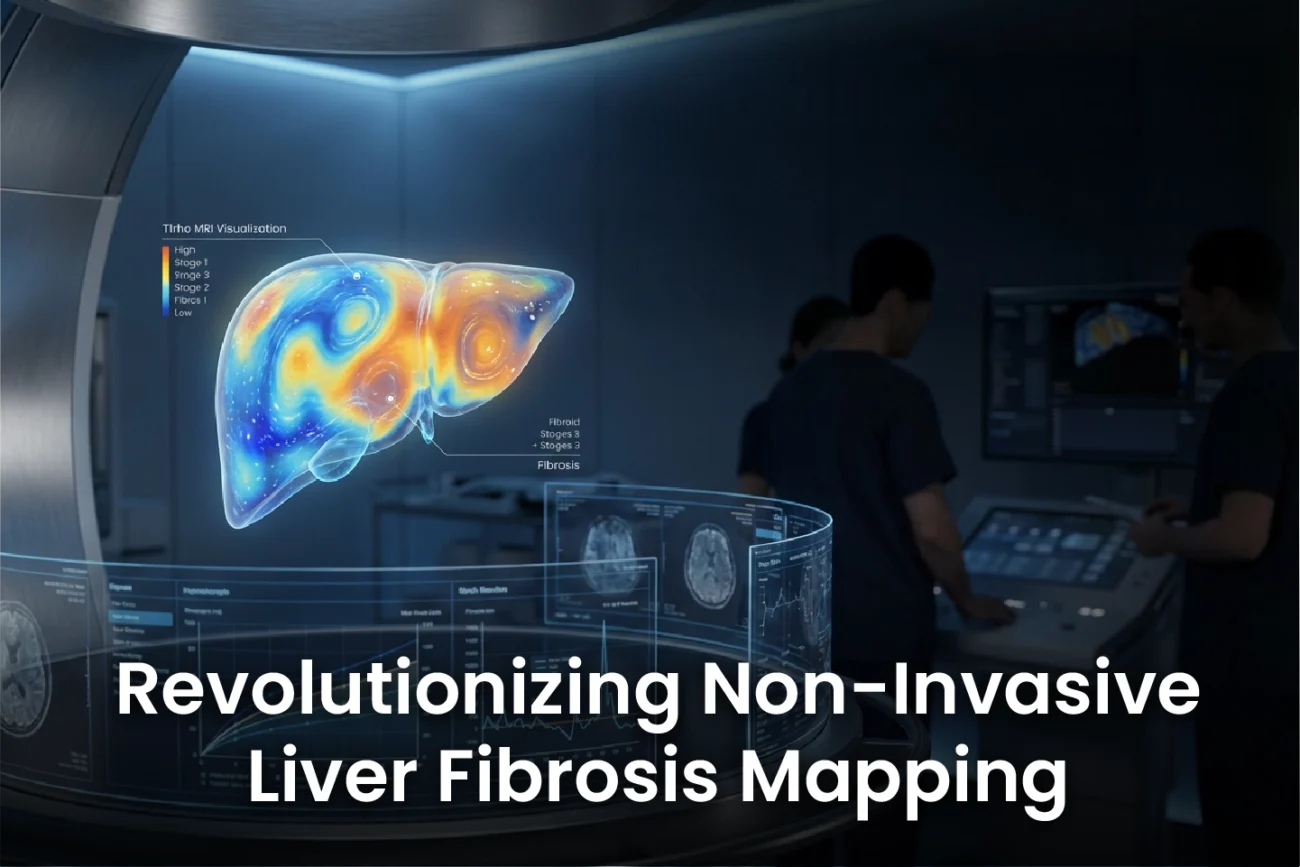
T1ρ liver fibrosis mapping is emerging as a powerful non-invasive tool for diagnosing significant hepatic fibrosis in patients with chronic liver disease (CLD). New clinical data demonstrates the technique offers high diagnostic accuracy, which is comparable to that of established methods like Magnetic Resonance Elastography (MRE) and Extracellular Volume Fraction (ECV) mapping. This advancement holds significant promise. It provides clinicians with a safe and highly precise alternative to invasive liver biopsy for staging fibrosis.
Understanding T1ρ Mapping and Study Design
The recent prospective clinical study included 112 participants with CLD. Notably, 53 participants also presented with hepatic steatosis (fatty liver disease). Researchers conducted a comprehensive liver Magnetic Resonance Imaging (MRI) protocol. Consequently, this included T1ρ mapping, MRE, Proton Density Fat Fraction (PDFF), and ECV measurements. The study’s main goal was to evaluate T1ρ mapping’s diagnostic performance. Therefore, they used MRE-based liver stiffness as the non-invasive reference standard for significant fibrosis (defined as ≥F2).
For individuals without steatosis (PDFF ≤ 5%), the significant fibrosis threshold was set at 3.66 kPa. Conversely, for participants with hepatic steatosis (PDFF > 5%), the threshold was slightly lower at 3.14 kPa. This design acknowledges that liver fat may affect stiffness measurements.
High Diagnostic Accuracy for Significant Fibrosis
The results decisively showed that all assessed quantitative mapping parameters were significantly elevated in participants with significant fibrosis (F2 or higher). Specifically, the mean T1ρ value for the significant fibrosis group was 110 ± 15 ms. This contrasts sharply with the mean T1ρ value of 92 ± 6 ms in the non-fibrotic group (p < 0.001). This large difference confirms T1ρ’s sensitivity to fibrotic changes.
Furthermore, the diagnostic performance of T1ρ mapping was excellent. The technique achieved an Area Under the Curve (AUC) of 0.93 for detecting significant fibrosis (≥F2). This impressive result suggests T1ρ mapping is a reliable method. It performs comparably to ECV (AUC 0.94) and MRE (AUC 0.96). Consequently, T1ρ mapping provides a valuable non-invasive option for accurate disease staging in CLD patients.
T1ρ Mapping: The Edge in Liver Fibrosis Assessment
The study provides robust clinical validation for T1ρ mapping. This makes it a strong candidate for routine clinical use alongside other multiparametric MRI techniques. Moreover, the finding that T1ρ values are significantly increased in fibrosis, independent of the presence of hepatic steatosis, is highly important. Because fatty liver disease (SLD) is a global epidemic, a reliable non-invasive tool that is not confounded by fat is urgently needed. T1ρ mapping appears to fill this need effectively. Therefore, clinicians can now rely on this metric to differentiate between simple steatosis and steatohepatitis with accompanying fibrosis.
Frequently Asked Questions
Q1: What is the clinical significance of T1ρ mapping’s high AUC value?
An Area Under the Curve (AUC) of 0.93 indicates excellent diagnostic accuracy. It means the T1ρ mapping technique can correctly differentiate between patients with and without significant liver fibrosis (≥F2) 93% of the time, which is highly reliable for clinical decision-making.
Q2: How does T1ρ mapping perform in patients with fatty liver disease?
The study included a significant number of patients with hepatic steatosis (fatty liver disease). It showed that T1ρ mapping remains highly effective and accurate for fibrosis staging even in the presence of liver fat (SLD). This makes it a particularly valuable tool for a wide range of chronic liver diseases.
Q3: Why is a non-invasive tool like T1ρ mapping important for liver fibrosis?
Liver biopsy, the traditional gold standard, is invasive, carries risks, and suffers from sampling variability. Non-invasive tools like T1ρ mapping offer a safe, repeatable, and objective way to stage liver fibrosis, helping to monitor disease progression and treatment response without procedural complications.
References
- Mesropyan N et al. Clinical validation of T1ρ mapping for the assessment of hepatic fibrosis in patients with chronic liver disease. Eur Radiol. 2025 Dec 20. doi: 10.1007/s00330-025-12225-5. PMID: 41420707.
- Xie S et al. Impact of Liver Fibrosis and Fatty Liver on T1rho Measurements: A Prospective Study. Korean J Radiol. 2017 Nov-Dec.
- Wang YX. Evaluation of liver fibrosis with T1ρ MR imaging. Quant Imaging Med Surg. 2013 Aug.
- Retrospective study on MRI accuracy in assessing liver fibrosis among patients with chronic liver disease – Bioinformation. 2025.