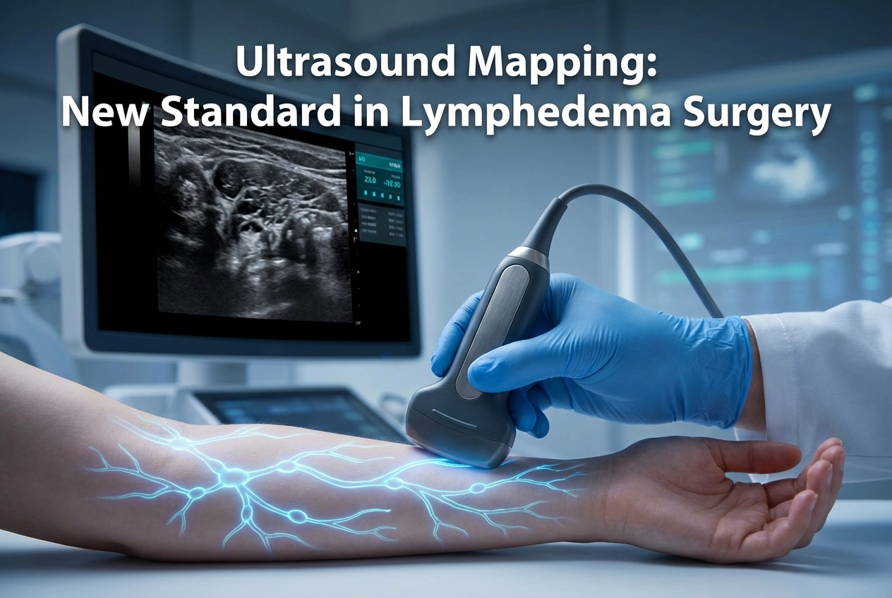
Lymphatic-venous anastomosis (LVA) is a highly effective surgical treatment for chronic lymphedema, yet its success fundamentally relies on accurately identifying patent lymphatic vessels. Unfortunately, the current standard, Indocyanine Green (ICG) lymphography, cannot always successfully map these crucial vessels. Therefore, surgeons increasingly need a more reliable preoperative method. A recent study exploring the combination of High-Frequency Ultrasound (HFUS) and Contrast-Enhanced Ultrasound (CEUS) confirms that Ultrasound Lymphatic Mapping provides a powerful, reliable alternative when ICG lymphography is not feasible. Furthermore, the combined approach offers superior visualization and mapping accuracy.
Ultrasound Lymphatic Mapping: The Combined Approach
Researchers conducted a study on 111 lymphedema patients who showed a non-linear pattern on ICG lymphography, indicating poor visualization. They utilized combined HFUS and CEUS for preoperative lymphatic mapping. The study successfully identified 345 lymphatics in the limbs and 52 in the perineum for anastomosis. For instance, measurements showed a lymphatic vessel inner diameter of 0.5–0.9 mm and a depth of 9–10 mm. Significantly, the combined HFUS plus CEUS technique dramatically improved detection sensitivity compared to either modality alone. For example, HFUS alone identified 114 vessels with 88.6% accuracy, and CEUS alone identified 22 vessels with 90.9% accuracy. Conversely, the combined approach successfully identified 313 vessels, achieving an impressive 91.1% accuracy. Consequently, this superior detection leads to better surgical targeting. Surgeons successfully performed the planned LVA procedures based on the ultrasound mapping results.
Improved Patient Outcomes with US-Guided LVA
The study further evaluated patient outcomes by comparing pre- and post-operative measurements. The LVA surgery, based on combined Ultrasound Lymphatic Mapping achieved a significant reduction in limb swelling. For instance, the average limb circumference decreased from 39.3 ± 7.4 cm to 37.8 ± 7.1 cm. Furthermore, the average limb volume also showed a statistically significant reduction, moving from 81.1 ± 35.8 L to 74.2 ± 33.4 L. This evidence strongly supports the use of combined HFUS and CEUS as a robust, non-invasive method for identifying target vessels and improving surgical results in complex lymphedema cases. Additionally, other research also highlights the advantages of Ultra-High-Frequency Ultrasound (UHFUS) in depicting micro-vessels as small as 250 µm and overcoming the depth and resolution limitations of ICG lymphography. Ultimately, the high success rate of LVA surgery, when guided by precise ultrasound mapping, confirms its essential role in modern lymphedema treatment.
Frequently Asked Questions
Q1: Why is High-Frequency Ultrasound (HFUS) needed if ICG lymphography is the standard?
ICG lymphography has limitations, including poor resolution and limited tissue penetration (about 2 cm). Importantly, in advanced lymphedema, dermal backflow of the dye can obscure the deeper, functional lymphatic vessels. HFUS, especially when combined with CEUS and microbubbles, overcomes these issues, offering superior visualization of the tiny vessels (less than 1 mm) required for LVA.
Q2: How accurate is the combined HFUS + CEUS technique for lymphatic mapping?
The study showed that the combined HFUS + CEUS approach accurately identified vessels for LVA with a detection sensitivity of 91.1%. This was significantly higher than either HFUS or CEUS used individually. This superior accuracy helps surgeons precisely target vessels, directly contributing to the high success rate of LVA procedures.
Q3: What post-operative improvements did patients experience with this ultrasound-guided surgery?
Patients experienced clinically significant reductions in limb swelling following LVA surgery guided by this technique. The study noted a reduction in average limb circumference from 39.3 cm to 37.8 cm, alongside a statistically significant reduction in limb volume.
References
- Yuan S et al. High-frequency ultrasound combined with microbubbles for preoperative lymphatic mapping for lymphedema with a non-linear pattern in indocyanine green lymphography. Eur Radiol. 2026 Jan 24. doi: 10.1007/s00330-025-12293-7. PMID: 41579179.
- Boutros S et al. Ultra-high-frequency ultrasound in lymphatic surgery: the next frontier. Microsurgery. 2024 Oct 25. doi: 10.1002/micr.31195.
- Suami H et al. Lymphatic Mapping with Contrast-enhanced Ultrasound for Lymphaticovenous Anastomosis Surgery: How We Do It. Plast Reconstr Surg Glob Open. 2023 Oct 12;11(10):e5328.
- Chang DW et al. Noncontrast Ultra-High-Frequency Ultrasound for Preoperative Lymphovenous Mapping in Patients Undergoing Lymphaticovenous Anastomosis Surgery for Extremity Lymphedema. AJR Am J Roentgenol. 2024 Jan;222(1):e23307.