Evaluating adnexal masses accurately remains a critical challenge for gynecologists worldwide. Modern technology now offers ultrasound radiomics models to assist in distinguishing between benign and malignant tumors. A recent study explored these machine-learning tools to improve diagnostic precision. Researchers compared a new clinical-radiomics model against the established Assessment of Different NEoplasias in the adneXa (ADNEX) model.
The retrospective study analyzed data from over 2,000 patients at a major Italian hospital. Each patient underwent ultrasound imaging before surgical removal and histological confirmation. Specifically, the team focused on images from the International Ovarian Tumor Analysis database. They used the eXtreme Gradient Boosting algorithm to build a model incorporating patient age and CA 125 levels. Additionally, they selected 14 specific radiomics features to enhance prediction accuracy.
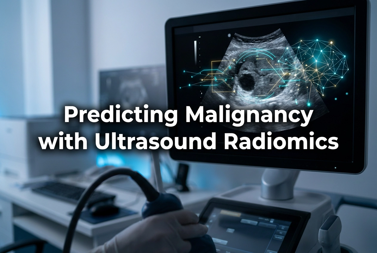
Utility of Ultrasound Radiomics Models
The results showed that the radiomics approach achieved a respectable AUC of 0.89. However, the traditional ADNEX model outperformed it with an AUC of 0.95. At a 10% risk threshold, the ADNEX model demonstrated superior sensitivity and specificity. Consequently, clinicians should still rely on the ADNEX model for current practice. Nevertheless, the study highlights the potential for AI to support clinical decisions in the future.
Furthermore, the researchers identified several areas for improvement. Future studies should include multiple images per tumor to capture more comprehensive data. Additionally, external validation across different centers will ensure the generalizability of these tools. Therefore, while AI shows promise, traditional clinical models remain the gold standard for now. Practitioners should stay informed about these technological advancements as they evolve.
Frequently Asked Questions
Q1: How did the radiomics model perform compared to the ADNEX model?
The ADNEX model performed significantly better than the radiomics model in this study. It achieved an AUC of 0.95, while the radiomics model reached 0.89 at its highest performance.
Q2: What features did the machine-learning model use for its predictions?
The model used patient age, serum CA 125 levels, and 14 distinct radiomics features extracted from digital ultrasound images.
Q3: Will these models replace current clinical methods?
Not yet. While the results are promising, the study suggests that the ADNEX model remains more effective for distinguishing between benign and malignant masses in clinical settings.
References
- Moro F et al. Developing and validating ultrasound-based machine-learning models incorporating radiomics features to predict malignancy in adnexal masses. Ultrasound Obstet Gynecol. 2026 Mar 30. doi: 10.1002/uog.70203. PMID: 41906961.
- Van Calster B et al. Evaluating the risk of ovarian cancer before surgery using the ADNEX model to differentiate between benign, borderline, early stage invasive, advanced stage invasive, and metastatic tumors: multicentre external validation study. BMJ. 2014;349:g5920.
- Timmerman D et al. Simple ultrasound-based rules for the diagnosis of ovarian cancer. Ultrasound Obstet Gynecol. 2008;31(6):681-90.