AI in ARIA Detection: Expert Safety Guide for Clinicians
Amyloid-targeting monoclonal antibodies have inaugurated an exciting era in the care of early Alzheimer disease. However, these novel disease-modifying agents bring substantial clinical safety challenges. Specifically, amyloid-related imaging abnormalities represent a major complication requiring rigorous surveillance. Accurate ARIA detection on MRI remains paramount because imaging findings directly dictate treatment continuation, temporary suspension, or dosage adjustments. As clinics adopt these therapies, radiology teams face increasing scan volumes and interreader variability. Therefore, clinical artificial intelligence systems have emerged to provide critical diagnostic assistance.
Understanding ARIA and Current Surveillance Challenges
Anti-amyloid therapies cause two distinct types of imaging changes. First, ARIA-E manifests as vasogenic edema or sulcal effusions within cortical and subcortical regions. Second, ARIA-H involves cerebral microhemorrhages and superficial cortical siderosis. Most patients remain completely asymptomatic during these events. Nevertheless, severe cases can produce significant focal deficits, headaches, encephalopathy, or seizures. Consequently, regulatory protocols mandate serial MRI evaluations before initiation and during early therapeutic cycles.
Interpreting these surveillance scans presents notable practical obstacles. Subtle swelling or tiny punctate hemorrhages frequently elude visual inspection on routine sequences. Furthermore, high interreader variability persists across practice environments, especially in community facilities. General radiologists often review these specialized scans without subspecialty neuroradiology consultation. In addition, growing patient cohorts substantially elevate overall imaging volume. Thus, clinical services require robust mechanisms to ensure consistent diagnostic accuracy.
Optimizing ARIA Detection on MRI with AI Workflows
An expert multidisciplinary panel recently reviewed the implementation of artificial intelligence decision-support platforms for ARIA surveillance. The panel affirmed that AI models improve lesion sensitivity, notably for small microbleeds and focal cortical edema. However, algorithms also generate false-positive markings that necessitate careful human review. Therefore, specialists advocate a strict "radiologist-in-the-loop" framework rather than autonomous evaluation.
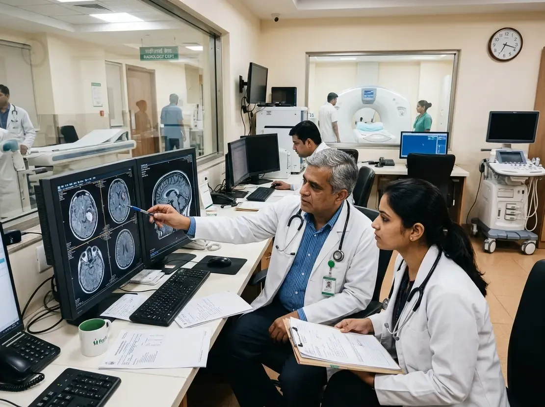
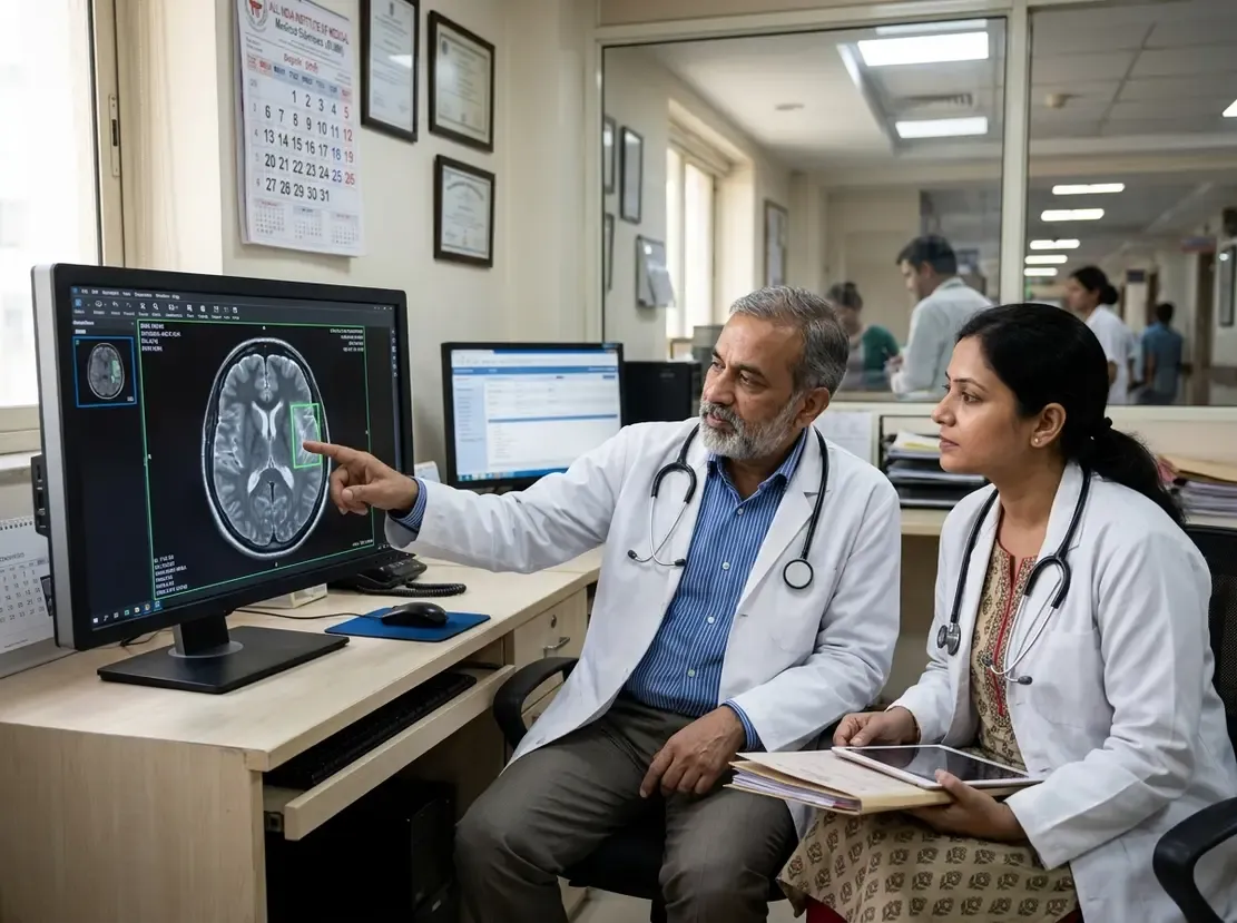
In this workflow, the radiologist conducts an initial independent review of standard MRI sequences. Next, the physician activates the AI overlay to scrutinize computer-flagged regions of interest. Moreover, the software measures lesion numbers, tracks longitudinal volumetric changes, and grades severity against clinical criteria. This collaborative approach enhances diagnostic consistency while preserving clinical oversight. Consequently, treating neurologists gain reliable data to make safe prescribing decisions.
Technical Discrepancies and Safe Implementation Standards
Commercially available AI software solutions exhibit noticeable discrepancies across clinical settings. For example, commercial packages differ significantly in technical capabilities, regulatory clearances, and validation cohorts. Many tools trained on selective clinical trial data struggle with heterogeneous real-world scanner hardware. Furthermore, variations in slice thickness and pulse sequences can degrade algorithmic precision.
To guarantee patient safety, hospitals must establish rigorous quality assurance protocols. First, imaging facilities must harmonize acquisition parameters, such as 3D T2-FLAIR and susceptibility-weighted imaging sequences. Second, clinical departments must implement continuous prospective monitoring to evaluate local model reliability. In addition, institutions should track false-positive rates to prevent unnecessary treatment interruptions. Ultimately, multidisciplinary collaboration between neurologists and specialists trained through an advanced clinical program in neuroradiology ensures responsible technology adoption.
Frequently Asked Questions
Q1: What is the primary safety concern with anti-amyloid therapies in Alzheimer disease?
The primary safety concern involves amyloid-related imaging abnormalities, divided into ARIA-E (brain edema or effusion) and ARIA-H (microhemorrhages or superficial siderosis). While many cases present without symptoms, severe events can cause significant neurological complications that necessitate dose adjustments or treatment suspension.
Q2: Can artificial intelligence replace radiologists in ARIA surveillance?
No, artificial intelligence cannot replace human radiologists. Expert panels recommend a radiologist-in-the-loop framework where AI functions strictly as a decision-support overlay. This safeguards against false-positive flags and maintains expert physician judgment in diagnostic interpretation.
Q3: Why is sequence harmonization necessary for AI-based ARIA evaluation?
AI algorithms demonstrate sensitivity to variations in magnetic field strength, slice thickness, and pulse sequences like FLAIR and SWI. Therefore, standardized imaging protocols across MRI scanners ensure reproducible algorithmic performance and reduce technical artifacts.
References
- Petrella JR et al. Artificial Intelligence-Based Detection of ARIA on MRI During Alzheimer Disease Therapy: Expert Opinion on Responsible Clinical Integration. AJR Am J Roentgenol. 2026 Sep 16. doi: 10.2214/AJR.26.35514. PMID: 42747213.
- Petrella JR, Liu AJ, Wang LA, Doraiswamy PM. Artificial Intelligence–Assisted Detection of Amyloid-Related Imaging Abnormalities: Promise and Pitfalls. AJNR Am J Neuroradiol. 2026 Feb; doi: 10.3174/ajnr.A8946.
- Christodoulou R, Papanastasiou E, Hadjigeorgiou GM. From Lesion to Decision: AI for ARIA Detection and Predictive Imaging in Alzheimer's Disease. Biomedicines. 2025 Nov;13(11):2739.