Management of **B3 breast lesions** presents a significant challenge for multidisciplinary teams worldwide, including in India. These high-risk lesions, which specialists diagnose via image-guided biopsy, carry an uncertain malignant potential. Consequently, many patients historically undergo surgical excision to definitively rule out carcinoma. Therefore, a recent systematic review and meta-analysis investigates whether contrast-enhanced breast MRI (CE-MRI) offers sufficient negative predictive value (NPV) to safely identify lesions clinicians can manage with surveillance alone, thus avoiding unnecessary operations.
The study sought to determine the added diagnostic value of CE-MRI in patients who have a high-risk (B3) lesion following an image-guided biopsy. Researchers performed a systematic review of eligible English-language articles. Ultimately, they calculated key diagnostic metrics like sensitivity, specificity, and likelihood ratios. Furthermore, the analysis utilized a bivariate random-effects model for robust results. **However**, the most clinically relevant finding centered on the post-test probability of a negative MRI result.
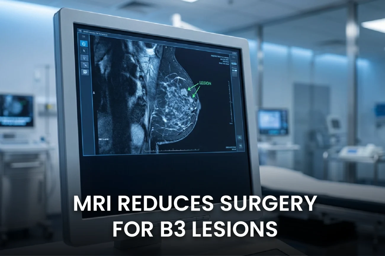
Maximizing Non-Surgical Management for B3 Breast Lesions
To safely manage a lesion with follow-up rather than surgery, clinicians must establish a low threshold for residual malignancy. The widely accepted threshold for downgrading a BI-RADS 4 lesion to BI-RADS 3 is a less than 2% probability of malignancy. **Therefore**, the authors employed Fagan nomograms to identify the maximum pre-test probability at which a negative CE-MRI could meet this 2% benchmark. This critical threshold essentially defines the highest initial risk level where a negative MRI can reassure a patient, helping them avoid a scalpel.
Clinicians know CE-MRI offers high sensitivity for detecting invasive and in situ breast cancer. When the imaging is negative, the resulting post-test probability of malignancy drops dramatically. **In fact**, the high NPV observed in similar studies suggests that a substantial number of B3 lesion patients could safely move from a recommendation for surgical excision to one for imaging surveillance. Minimally invasive techniques like Vacuum-Assisted Excision (VAE) and follow-up now constitute the preferred management for many B3 lesion subtypes in modern guidelines, particularly those without atypia. **Moreover**, Indian best practice guidelines also advocate for a standardized, holistic approach to breast care and support the use of MRI in high-risk settings. Consequently, this meta-analysis provides strong evidence to integrate CE-MRI into multidisciplinary decision-making pathways. This ultimately helps reduce overtreatment and its associated complications.
Frequently Asked Questions
Q1: What is a B3 breast lesion?
B3 breast lesions are a heterogeneous group of findings on a core needle biopsy that are categorized as “lesions of uncertain malignant potential.” They are considered high-risk because they have a variable, but non-zero, chance of being upgraded to in situ or invasive carcinoma upon surgical excision, often ranging from 10% to 35% depending on the specific subtype and the presence of atypia.
Q2: Why is Contrast-Enhanced MRI (CE-MRI) useful for B3 lesions?
CE-MRI has very high sensitivity for detecting breast malignancy. Therefore, when a CE-MRI result is negative (i.e., it shows no suspicious enhancement), its high Negative Predictive Value (NPV) can effectively “rule out” malignancy. This allows a significant number of patients to safely avoid surgical excision and opt for less invasive management, like imaging follow-up.
Q3: How does the “2% malignancy threshold” relate to avoiding surgery?
The 2% threshold is a widely accepted risk level in breast imaging, corresponding to downgrading a lesion from BI-RADS 4 (Suspicious) to BI-RADS 3 (Probably Benign). The study used this threshold to determine the maximum initial risk (pre-test probability) at which a patient, after a negative CE-MRI, would fall below the 2% risk, confirming they are safe for imaging surveillance rather than surgery.
References
- Vatteroni G et al. Potential role of breast MRI to identify patients with high-risk lesions who might avoid surgery: a systematic review and meta-analysis. Eur Radiol. 2026 Jan 17. doi: 10.1007/s00330-025-12291-9. PMID: 41545805.
- Schulze S, et al. B3 Lesions: Radiological Assessment and Multi-Disciplinary Aspects. *Breast Care*. 2020;15(6):534-541.
- Preibsch H, et al. Malignancy rates of B3-lesions in breast magnetic resonance imaging – do all lesions have to be excised? *BMC Med Imaging*. 2018;18(1):33.
- Breast Imaging Society, India. Best Practice Guidelines for Breast Imaging, Breast Imaging Society, India: Part-2. *Annals of the National Academy of Medical Sciences (India)*. 2023;59(3):144-159.