Closed Spinal Dysraphism: Fetal Signs to Child Outcomes
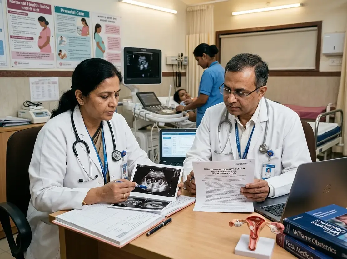
Clinicians frequently evaluate complex fetal anomalies during routine anomaly scans in obstetric practice.
Therefore, accurate characterization of spinal defects plays a vital role in parental counseling.
Closed spinal dysraphism encompasses skin-covered congenital spinal lesions that differ substantially from open neural tube defects.
In addition, these lesions display a broad spectrum of structural severity and long-term functional implications.
Recent ten-year multicenter evidence offers clear guidance regarding prenatal sonographic accuracy and postnatal outcomes.
Understanding Closed Spinal Dysraphism Classification
Historically, varying terminology created confusion among fetal medicine specialists and pediatric neurosurgeons.
However, the updated Orphanet consensus framework provides a standardized diagnostic system for clinical practice.
Saccular anomalies account for more than eighty-five percent of prenatally diagnosed closed lesions.
Specifically, limited dorsal myeloschisis represents the most common saccular subtype identified during fetal evaluation.
Posterior meningocele and conus spinal cord lipoma constitute the remaining frequent presentations in this cohort.
Furthermore, most spinal defects localize to the lumbar or sacral spinal regions.
Associated cranial malformations occur in approximately seventeen percent of affected fetuses.
Consequently, clinicians must perform dedicated neurosonography to evaluate the entire neuraxis thoroughly.
Prenatal Diagnostic Concordance and Surgical Care
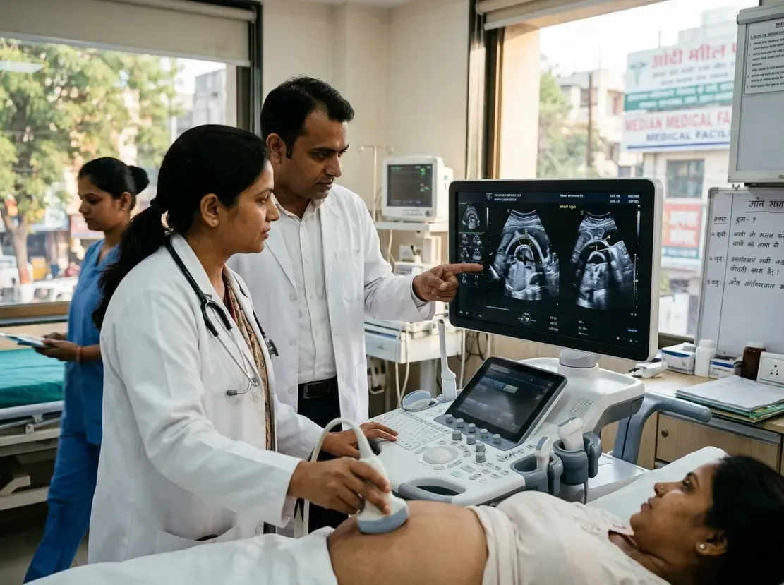
High-resolution prenatal ultrasound demonstrates exceptional diagnostic concordance with postnatal evaluations.
In fact, fetal ultrasound correctly identifies the specific anatomical subtype in nearly ninety percent of cases.
Minor adjustments to the initial diagnosis occur postnatally in approximately ten percent of neonates.
Nevertheless, prenatal imaging reliably informs delivery room management and early pediatric intervention.
After birth, pediatric neurosurgeons operate on more than ninety percent of symptomatic or tethered infants.
Timely surgery aims to release spinal cord tethering and prevent progressive neurological decline.
Therefore, fetal medicine units must coordinate delivery within tertiary hospitals equipped with pediatric neurosurgical teams.
Postnatal Functional Outcomes and Long-Term Morbidity
Long-term pediatric follow-up reveals reassuring functional motor capacity for most children.
For example, nearly ninety-five percent of children achieve independent ambulation by thirty-six months of age.
In contrast, urological dysfunction remains a prominent and persistent clinical challenge over time.
Bladder dysfunction affects nearly forty percent of children during early childhood follow-up assessments.
Gastrointestinal symptoms occur less frequently, impacting approximately twenty-two percent of these pediatric patients.
Because voiding dysfunction can cause silent renal deterioration, proactive urological monitoring remains essential.
Hence, routine urodynamic studies and renal ultrasonography should begin shortly after birth.
Clinical Considerations for Obstetricians in India
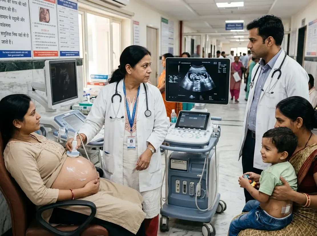
In India, routine mid-trimester ultrasound screening detects an increasing volume of occult spinal dysraphism.
Thus, fetal radiologists must carefully inspect the overlying cutaneous contour and intact skin layer.
When sonographers detect a closed lesion, they should search diligently for intracranial Chiari malformations.
Additionally, obstetricians should offer comprehensive counseling that distinguishes closed dysraphism from open spina bifida.
Open spina bifida carries substantial motor deficits, whereas closed lesions usually allow preserved ambulation.
However, couples must understand the ongoing necessity of postnatal neurosurgical and urological surveillance.
Frequently Asked Questions
Q1: How does closed spinal dysraphism differ from open spina bifida?
Closed spinal dysraphism features intact skin covering the underlying vertebral and neural defect. In contrast, open spina bifida involves exposed neural tissue without continuous cutaneous coverage, resulting in higher risks of motor deficit and Chiari II malformation.
Q2: What is the likelihood of independent walking in children with closed dysraphism?
Motor outcomes are generally very favorable for affected infants. In fact, research demonstrates that approximately ninety-four percent of children achieve independent ambulation by three years of age.
Q3: Why is early urological surveillance critical after birth?
Although motor function remains preserved in most patients, urinary dysfunction affects nearly forty percent of children. Therefore, early urodynamic testing prevents recurrent urinary tract infections and protects long-term renal function.
References
- Athiel Y et al. Prenatal findings and postnatal outcomes in cases of closed spinal dysraphism: 10-year two-center cohort study. Ultrasound Obstet Gynecol. 2026 Sep 20. doi: 10.1002/uog.70340. PMID: 42763785.
- Dhombres F, de Saint-Denis T, Thompson D, Mosiello G, et al. Revised Orphanet Nomenclature and Classification for Spina Bifida and Other Spinal Dysraphisms (SBoD). Orphanet J Rare Dis. 2025;20(1):145.
- Adler M, et al. Prenatal diagnosis and postnatal outcome of closed spinal dysraphism. Prenat Diagn. 2024;44(4):499-510.