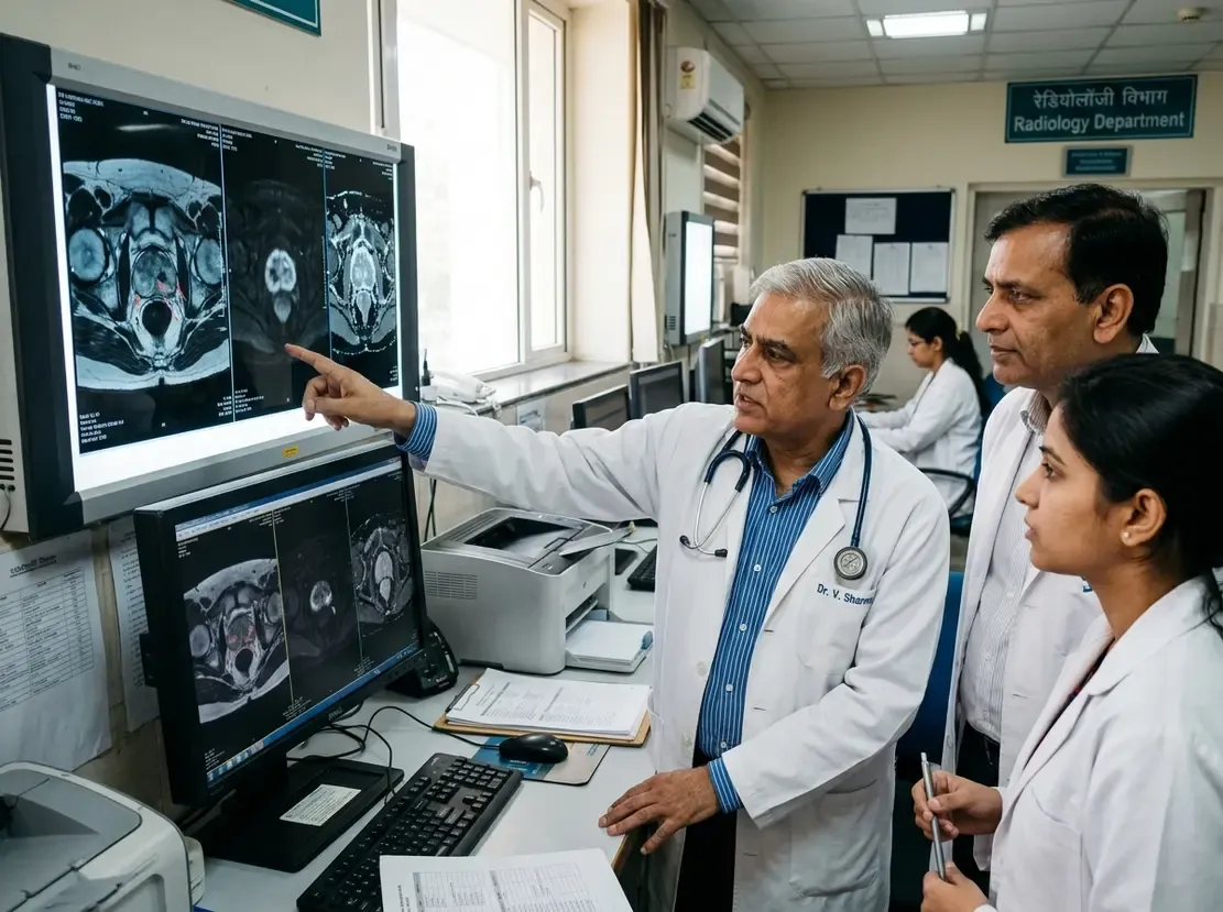
AI-Driven CT Anatomical Labelling Streamlines Imaging
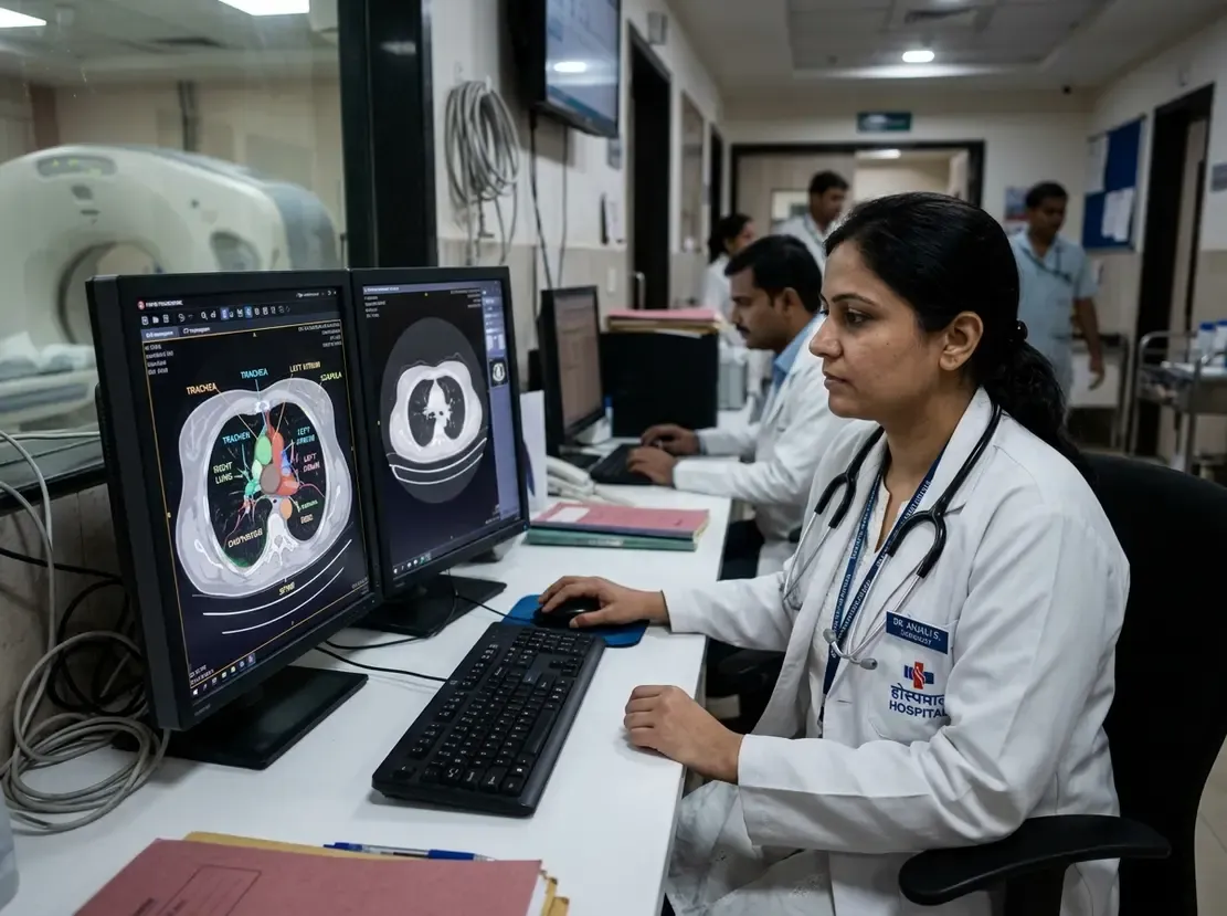
Radiologists frequently struggle with inconsistent series descriptions across diverse scanner vendors. Therefore, automated CT anatomical labelling offers a transformative solution for hospital imaging networks. Traditional data pipelines depend heavily on unstructured text tags within DICOM headers. Unfortunately, these textual entries often feature typos, varied terminology, or incomplete descriptions. Consequently, clinicians must manually verify series before feeding scans into artificial intelligence algorithms. A novel deep learning system now addresses this challenge directly.
Overcoming Metadata Challenges with CT Anatomical Labelling
The Rapid Analysis and Processing of Image Data framework, known as RAPID, redefines image indexing. Specifically, RAPID combines deep learning models with DICOM spatial geometry. Rather than trusting text tags, the architecture inspects 2D scout topograms. Researchers trained three separate YOLOv8-based neural networks on over 65,000 patient examinations. These dedicated models perform global anatomical classification, body-region detection, and precise landmark detection. Thus, the system extracts ground-truth anatomical coverage directly from imaging pixels.
In addition, this spatial approach bridges topogram coordinates with volumetric axial slices. As a result, PACS archives can automatically categorise chest, abdominal, and extremity examinations. Moreover, the automated pipeline prevents manual sorting errors during emergency triaging.
Robust Diagnostic Validation and Real-World Accuracy
The technical evaluation demonstrated outstanding predictive capabilities across internal and external cohorts. For instance, the global classification model reached an internal F1 score of 0.920. Furthermore, external validation yielded an impressive classification F1 score of 0.970. Body region detection achieved an internal mAP50 of 0.993, confirming exceptional localization reliability. Similarly, anatomical landmark detection produced an internal mAP50 of 0.958. Consequently, external testing maintained high precision across different patient demographics.
Beyond statistical benchmarks, three independent radiologists evaluated 150 randomly chosen detection outputs. Notably, expert clinical assessments closely mirrored the technical metrics. Inter-rater agreement confirmed that the YOLOv8 detections aligned with clinical expectations. Therefore, the framework proves ready for integration into busy hospital radiology suites.
Clinical Implications for Radiology Workflows
High-volume imaging centers in India face mounting burdens from uncurated imaging datasets. Inconsistent DICOM headers severely disrupt automated AI triage algorithms and PACS viewing hanging protocols. However, topogram-derived anatomical labeling completely bypasses textual inconsistencies. As a result, imaging departments can process complex multi-vendor scans without tedious manual sorting.
Furthermore, large research registries benefit substantially from standardized spatial labels. Data scientists can rapidly aggregate precise body regions for training downstream diagnostic algorithms. Ultimately, this scalable automation elevates radiology throughput and enhances diagnostic quality across institutional networks.
Frequently Asked Questions
Q1: Why is textual DICOM metadata unreliable for CT series classification?
Hospital networks employ diverse scanners with custom protocols. Consequently, series descriptions often contain non-standard terminology, typos, or blank fields, which leads to cataloging errors.
Q2: How does the RAPID framework identify anatomical regions?
The framework utilizes three YOLOv8 deep learning models trained on 2D CT topograms. Furthermore, it integrates DICOM spatial coordinates to map image pixels directly to anatomical regions.
Q3: How do radiologists benefit from automated series labelling?
Automated labelling ensures consistent PACS hanging protocols and seamless AI triage. Therefore, radiologists spend less time organizing series and can prioritize urgent diagnostic interpretations.
References
- Wen Y et al. Topogram-based anatomical labelling of CT series: anatomy-aware CT data processing using deep learning. Eur Radiol. 2026 Sep 03. doi: 10.1007/s00330-026-12830-y. PMID: 42690467.
- Chiang CH, Weng CL, Chiu HW. Automatic classification of medical image modality and anatomical location using convolutional neural network. PLoS One. 2021;16(6):e0253205. doi: 10.1371/journal.pone.0253205.