
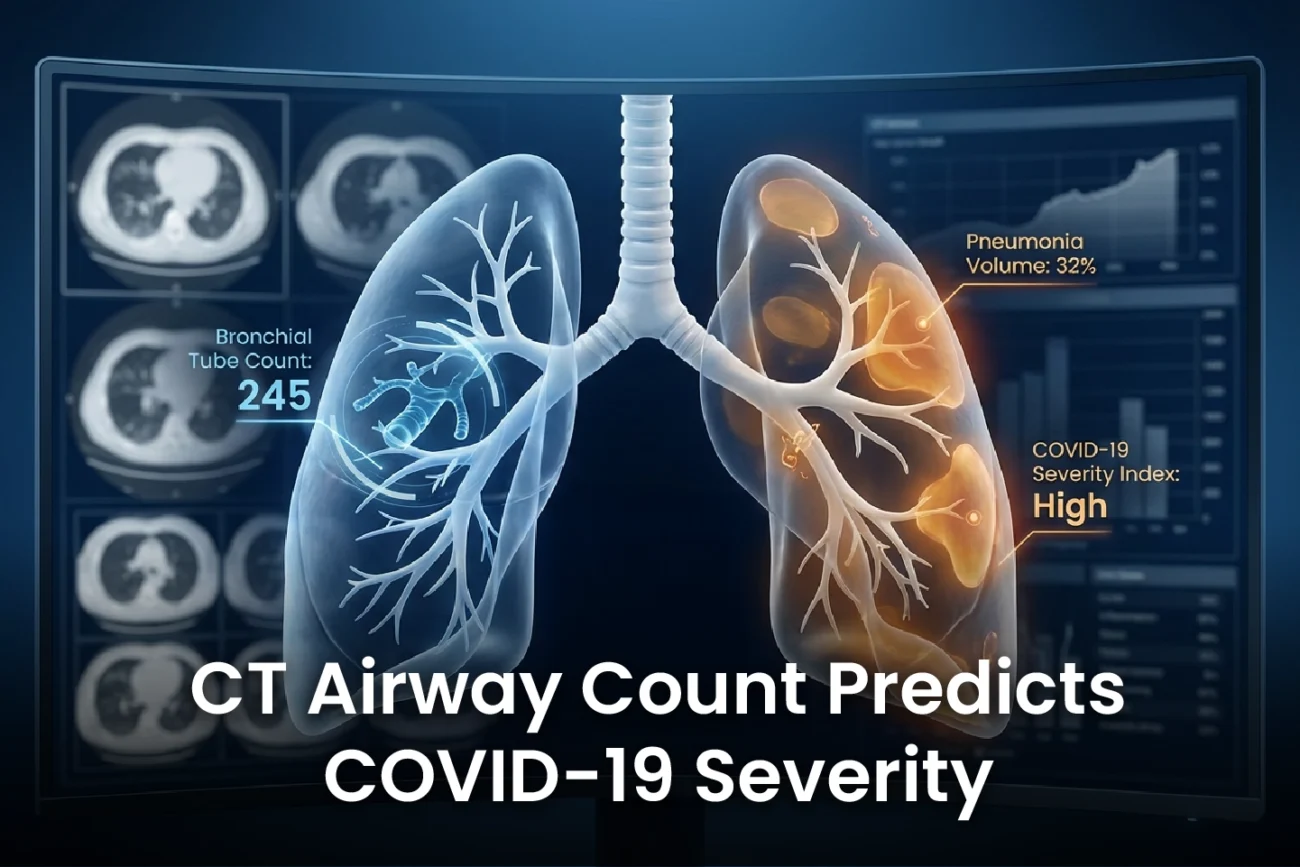
AI-driven analysis of chest computed tomography (CT) offers novel insights into COVID-19 prognosis. Integrated assessment of Total Airway Count (TAC) and pneumonia volume is emerging as a powerful CT prognostic marker for predicting critical illness in patients with coronavirus disease. However, the exact clinical relevance of CT-based airway tree structure remained unclear until recently. This new research provides clarity on how a combined metric can significantly improve patient risk stratification.
Study Findings on TAC and Pneumonia Volume
A multicenter retrospective cohort study in Japan examined 781 hospitalised COVID-19 patients. Researchers used artificial intelligence to segment both the airway tree and pneumonia regions. They measured the Total Airway Count (TAC) and the percentage of pneumonia volume in each patient. Additionally, the study focused on critical outcomes, which included the need for high-flow oxygen, invasive mechanical ventilation, or death. Critical outcomes were observed in 8.8% of all patients. Furthermore, these patients demonstrated a significantly higher TAC at admission.
The Role of the Integrated CT Prognostic Marker
Scientists divided the patients into four distinct groups using specific cutoff values (17.6% for pneumonia volume percent and 255 for TAC). Group D, characterised by both high TAC and high pneumonia volume, experienced the worst clinical course. In fact, this group showed the highest levels of inflammation and fibrosis markers, as well as the most complications. Consequently, Group D had a significantly higher risk of critical outcomes, even after adjusting for factors like age, sex, body mass index, total lung volume, and comorbidities.
Other research confirms that quantitative CT parameters, especially pneumonia volume, have high value for predicting patient clinical course and progression to critical illness in COVID-19 patients. Moreover, low CT-derived lung volume at admission also associates with poor clinical outcomes and post-hospitalization complications. Therefore, the integrated assessment combines two vital pieces of information to create a superior prognostic tool.
Clinical and Future Relevance
The combined TAC and pneumonia volume metric effectively predicted critical outcomes in the study population. This AI-driven integrated assessment has immediate clinical relevance for identifying high-risk patients early. In the 3-month follow-up, pneumonia volume improved in critical cases, but TAC did not. This longitudinal finding suggests that the initial anatomical changes (TAC) may persist in patients who require intensive care. Ultimately, this metric can potentially apply to various respiratory diseases, including infectious or interstitial pneumonia, expanding its utility beyond COVID-19.
Frequently Asked Questions
Q1: What are the two main metrics used in this integrated assessment?
The assessment combines Total Airway Count (TAC), which reflects the airway tree structure, and the percentage of pneumonia volume, both measured via AI segmentation on chest CT.
Q2: Which patient group had the worst outcomes?
Patients in Group D, defined by both a high Total Airway Count (TAC > 255) and a high pneumonia volume percent (> 17.6%), faced the highest risk of critical outcomes, including death or the need for mechanical ventilation.
Q3: Does the Total Airway Count (TAC) improve over time?
In the 3-month longitudinal analysis of critical cases, pneumonia volume improved, but the Total Airway Count (TAC) did not. This suggests that the initial TAC may represent a more persistent anatomical finding.
References
- Nakagawara K et al. Integrated assessment of total airway count and pneumonia volume on chest computed tomography as a prognostic biomarker for coronavirus disease. Eur Radiol. 2025 Dec 24. doi: 10.1007/s00330-025-12078-y. PMID: 41442001.
- Quantitative Evaluation of COVID-19 Pneumonia Lung Extension by Specific Software and Correlation with Patient Clinical Outcome. J Clin Med. 2021.
- CT Quantification of COVID-19 Pneumonia at Admission Can Predict Progression to Critical Illness: A Retrospective Multicenter Cohort Study. Acad Radiol. 2021.
- Kono T, et al. Lung volume measurement using chest CT in COVID-19 patients: a cohort study in Japan. Eur Radiol. 2024.