
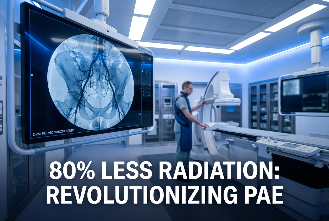
Prostatic Artery Embolization (PAE) offers a minimally invasive treatment for Benign Prostatic Hyperplasia (BPH). A prospective randomized trial now confirms that **Digital Variance Angiography (DVA)** can significantly reduce patient radiation exposure during this procedure. Consequently, this innovation addresses a critical safety concern in interventional radiology. The study demonstrated that an ultra-low-dose (ULD) DVA protocol successfully cut the stationary acquisition radiation dose by up to 80% compared to a standard Digital Subtraction Angiography (DSA) protocol.
DVA Revolutionizes PAE: Less Radiation, Same Quality
Study Design and Major Findings
Researchers enrolled 70 patients undergoing PAE and randomized them into two groups: a normal dose (ND) DSA group and an ultra-low dose (ULD) DSA group, with the latter utilizing DVA. Initially, the ULD protocol targeted a 72% detector dose reduction for stationary acquisitions. However, the protocol achieved an even greater reduction in stationary acquisition-related Dose-Area Product (DAP), reaching up to 80% (580 ± 66 vs 2872 ± 396 µGym²/patient, p < 0.001).
Maintaining diagnostic quality at such low doses is paramount. Therefore, the researchers compared the Contrast-to-Noise Ratios (CNR) and visual image quality. Importantly, DVA images consistently showed a significantly higher CNR than both the ND and ULD DSA images. Three blinded interventional radiologists evaluated vessel visualization and tissue blush using a 5-point Likert scale. Furthermore, DVA-supported ULD imaging proved non-inferior to ND DSA for visualizing large vessels and tissue blush. In contrast, the ULD group showed slightly, but significantly, lower scores for small vessel visualization. Nonetheless, the overall diagnostic utility remained acceptable.
Digital Variance Angiography: The New Standard for PAE?
The success of DVA in PAE builds upon earlier research showing its effectiveness in other procedures. For instance, DVA has demonstrated significant radiation exposure reduction in lower limb angiography, with stationary acquisition-related DAP reduced by over 60% while maintaining non-inferior image quality. This evidence base suggests DVA’s superior image processing algorithm, which enhances contrast and suppresses noise, offers a vital safety reserve. Moreover, the input study’s finding that the ultra-low dose approach works well in the technically demanding PAE procedure is a major advancement. This is crucial because minimizing radiation exposure for both the patient and the operating staff is a universal goal in interventional radiology. Consequently, the integration of DVA technology into PAE protocols promises a safer future for patients undergoing BPH treatment.
Frequently Asked Questions
Q1: What is the main benefit of Digital Variance Angiography (DVA) in PAE?
The primary benefit is a substantial reduction in patient radiation dose. The study showed that using an ultra-low-dose protocol with DVA can reduce the radiation dose-area product (DAP) during stationary acquisitions by up to 80% in Prostatic Artery Embolization (PAE).
Q2: Does the lower radiation dose affect image quality?
The lower radiation dose protocol, enabled by DVA, maintained diagnostic image quality. Specifically, it was non-inferior for visualizing large vessels and tissue blush, which are critical for the procedure. DVA images also demonstrated a significantly higher Contrast-to-Noise Ratio (CNR) than standard Digital Subtraction Angiography (DSA).
Q3: How does DVA compare to the standard DSA?
DVA utilizes an advanced motion-based algorithm to enhance contrast and suppress noise, giving it a “quality reserve” compared to the reference-standard DSA. This reserve allows interventionalists to drastically lower the radiation dose while preserving the necessary image quality for complex procedures like PAE.
References
- Alizadeh LS et al. Digital variance angiography enables up to 80% reduction in stationary acquisition radiation dose during prostatic artery embolization: a prospective randomized trial. Eur Radiol. 2026 Jan 03. doi: 10.1007/s00330-025-12235-3. PMID: 41483205.
- Digital variance angiography reduces radiation in PAE procedures. AuntMinnie. 2024 Nov 18.
- Radiation Exposure Reduction by Digital Variance Angiography in Lower Limb Angiography: A Randomized Controlled Trial. NIH. 2023 Apr 30.
- Digital variance angiography allows about 70% decrease of DSA-related radiation exposure in lower limb X-ray angiography. NIH. 2021 Nov 08.