Enhanced Myometrial Vascularity (EMV) is a common sonographic finding in the postpregnancy period, often seen after delivery, pregnancy termination, or pregnancy loss. Initially, this dynamic finding—which represents the involuting hypervascularity of the placental bed—was frequently mislabeled as an acquired uterine arteriovenous malformation (AVM). Consequently, this confusion led to unnecessary anxiety and inappropriate invasive treatments, like embolization or hysterectomy. Conversely, modern practice emphasizes distinguishing EMV from true AVMs to ensure fertility-preserving and appropriate management. Physicians must therefore understand the distinct physiology, imaging features, and clinical management pathways for Enhanced Myometrial Vascularity.
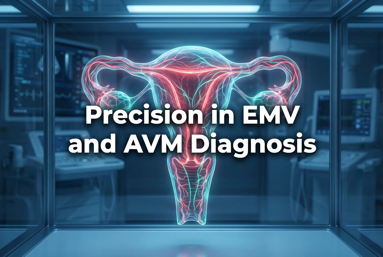
Enhanced Myometrial Vascularity: EMV vs. Uterine AVM
The distinction between Enhanced Myometrial Vascularity and a true uterine AVM is clinically critical. A congenital AVM is an extremely rare vascular malformation, which is generally not associated with pregnancy. Moreover, true AVMs do not regress spontaneously and are often difficult to treat, commonly requiring repeated embolization procedures. In contrast, EMV is a dynamic, acquired condition directly related to persistent or involuting uteroplacental circulation in the postpregnancy state. Specifically, EMV most often occurs in the presence of retained products of conception (RPOC). Importantly, most cases of EMV resolve spontaneously after the associated RPOC is expelled or removed. Mislabeling EMV as AVM can provoke overtreatment in patients who would otherwise experience this natural resolution.
Imaging Features and Differential Diagnosis
Color Doppler ultrasound (US) is the primary imaging modality for assessing these lesions. EMV is characterized on Doppler US by a tortuous, dilated myometrial vessel network exhibiting high-velocity, turbulent flow. Retained products of conception are a primary cause of persistent EMV. To aid differentiation, practitioners should note that the vascularity in RPOC-associated EMV commonly extends into the endometrium. However, true uterine AVMs principally involve only the myometrium. Although peak systolic velocity (PSV) on Doppler can show significant overlap between benign and pathological conditions, it is still used to stratify risk. A PSV measurement is vital for guiding therapeutic choices. Consequently, an accurate interpretation prevents the high risk of major hemorrhage often incorrectly associated with managing EMV via hysteroscopy or D&C.
Clinical Management of Enhanced Myometrial Vascularity
Management must always be guided by the patient’s clinical presentation, rather than solely relying on imaging results. For asymptomatic patients or those experiencing weak bleeding and low PSV (e.g., below 40 cm/s), a conservative approach is usually implemented. This typically involves expectant management with serial US scans until resolution. In fact, recent evidence supports a more conservative approach for EMV associated with RPOC, as the risk of major hemorrhage is lower than previously suspected. Conversely, in cases of significant vaginal bleeding and/or very high PSV (e.g., above 60–70 cm/s), interventional procedures like uterine artery embolization (UAE) are strongly suggested. Therefore, radiologists and clinicians must collaborate closely to customize treatment plans and ultimately prevent unnecessary interventions or even hysterectomy.
Frequently Asked Questions
Q1: What is the primary difference between EMV and AVM?
Enhanced Myometrial Vascularity (EMV) is an acquired, transient finding that occurs after a pregnancy. It is related to placental site changes and often resolves spontaneously. Conversely, a true Arteriovenous Malformation (AVM) is a rare, congenital vascular anomaly that does not spontaneously regress and is not associated with pregnancy.
Q2: How does the presence of Enhanced Myometrial Vascularity impact a surgical procedure?
Historically, EMV was mislabeled as AVM, which suggested a high risk of life-threatening bleeding with surgical procedures like D&C. However, the current understanding is that hysteroscopic removal of retained products of conception (RPOC) associated with EMV is generally safe, provided appropriate precautions are taken. This understanding avoids overtreatment with interventional radiology procedures.
Q3: How is Enhanced Myometrial Vascularity managed?
Management is patient-specific and depends on clinical symptoms and Doppler ultrasound findings. Asymptomatic patients or those with low flow velocities often undergo conservative, expectant management with follow-up scans. However, patients with heavy, persistent bleeding or very high peak systolic velocities (PSV) may require uterine artery embolization.
References
- Zamboni CG et al. Enhanced Myometrial Vascularity: Is It an Arteriovenous Malformation? Review of Definitions, Imaging Findings, and Management. Radiographics. 2026 Feb undefined. doi: 10.1148/rg.250152. PMID: 41610036.
- Understanding Enhanced Myometrial Vascularity. Empowered Women’s Health. 2022 Sep 20.
- Enhanced Myometrial Vascularity: A Case Report and Diagnostic Insights. eurekaselect.com. 2025 Mar 4.
- Uterine Vascular Abnormalities (AVM congenital and Acquired); Teaching Point for Radiologist – Iranian Congress of Radiology. 2023 Sep 18.
- About uterine enhanced myometrial vascularity: Doppler ultrasound could reduce misdiagnosed life-threatening vaginal bleeding after pregnancy and guide the management. NIH. 2022 Oct 25.
- Role of Ultrasonography in the Evaluation of Retained Products of Conception. thieme-connect.com. 2024 Jun 18.
- Enhanced myometrial vascularity on MRI in a patient with retained products of conception. nih.gov. 2026 Jan 12.
- Invited Commentary: Refining Diagnosis and Management of Enhanced Myometrial Vascularity versus Uterine Arteriovenous Malformation. RSNA Journals. 2026 Jan 29.
- Enhanced Myometrial Vascularity: Case Presentation and Review – YouTube. 2021 Jul 1.