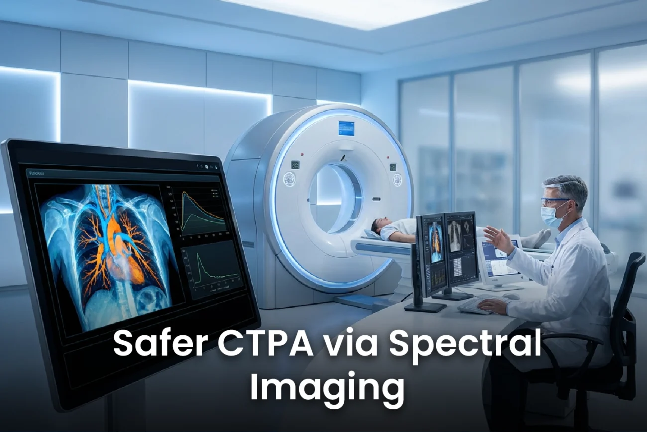
Computed Tomography Pulmonary Angiography (CTPA) is the gold standard for diagnosing pulmonary embolism (PE). However, the use of iodinated contrast media (ICM) poses risks, particularly for patients with renal impairment. A recent randomized controlled trial evaluated the efficacy of Low Contrast CTPA using a dual-layer detector spectral CT system with three different ICM protocols. The study demonstrated that using spectral imaging technology can maintain diagnostic image quality even with significantly reduced contrast volumes. Consequently, this supports safer and more efficient imaging practices for PE diagnosis.
Achieving Diagnostic Quality with Low Contrast CTPA Protocols
The randomized trial included 50 patients in each of the three contrast protocols (A: 40mL, B: 30mL, C: 20mL diluted). Conventional CT images showed that pulmonary artery vascular attenuation (VA) was above 200 Hounsfield Units (HU) in over 90% of cases for protocols A and B. However, the conventional VA dropped to only 70% in the lowest dose protocol C. Spectral CT technology effectively compensates for this decrease. The key finding shows that using Low-Energy Virtual Monoenergetic Images (LEVMI) significantly increased VA across all protocols. Furthermore, the minimum VA value observed in the LEVMI was 269 HU, which is a diagnostic level. This superior enhancement ensures reliable detection of PE. Consequently, the use of LEVMI provides diagnostic VA levels in the pulmonary arteries for all three administration protocols.
Spectral CT: The Mechanism of Contrast Dose Reduction
Dual-energy spectral CT uses advanced technology to generate virtual monoenergetic images. Therefore, radiologists can reconstruct these images at lower energy levels, typically between 50 and 70 keV, where iodine’s attenuation properties are naturally enhanced. This technical advantage boosts the visible contrast within the pulmonary vasculature. In the study, protocol C (20mL ICM) presented the lowest volumetric lung iodine density (VID) and the worst quality of the iodine map. However, it still successfully detected perfusion defects in 100% of the pulmonary embolism cases. Similarly, previous research confirms that DECT can safely reduce contrast dose by up to 25% without compromising image quality, which is especially important for patients with thyroid or renal issues.
Clinical Implications for Safer PE Imaging
The ability to achieve diagnostic quality with a drastically reduced contrast volume has significant clinical relevance. Reducing the dose of iodinated contrast media minimizes the risk of contrast-induced nephropathy (CIN), a major concern, particularly in elderly patients or those with pre-existing renal dysfunction. Using low-kVp or dual-energy protocols, other studies have successfully reduced contrast volume by 25% to 67% compared to standard doses. Moreover, the results from the randomized trial suggest that spectral CTPA protocols can be optimized for both safety and efficiency, supporting their integration into routine clinical practice.
Frequently Asked Questions
Q1: Why is reducing contrast dose for CTPA important?
Reducing the dose of iodinated contrast media (ICM) is clinically critical for patient safety. It lowers the risk of post-contrast acute kidney injury (PC-AKI) or contrast-induced nephropathy (CIN), a serious complication especially in patients who have pre-existing kidney problems. Furthermore, a reduced dose supports more efficient and cost-effective imaging protocols.
Q2: What is the main finding regarding the lowest contrast protocol (20mL)?
The lowest dose protocol (20mL ICM diluted with 20mL saline) provided sufficient image quality for diagnosis. Although its conventional CT images and iodine maps were lower quality, the Low-Energy Virtual Monoenergetic Images (LEVMI) achieved a diagnostic vascular attenuation in the pulmonary arteries. Significantly, this protocol detected perfusion defects in all pulmonary embolism cases.
Q3: How does spectral CT technology allow for contrast dose reduction?
Spectral CT, also known as dual-energy CT, allows the creation of virtual monoenergetic images (VMI) at low energy levels (e.g., 50-70 keV). The iodine contrast agent naturally shows higher attenuation (brightness) at these low energy levels, effectively boosting the signal and contrast-to-noise ratio in the pulmonary arteries. Consequently, this spectral boost allows the use of a lower volume of contrast agent while maintaining diagnostic image quality.
References
- Ferrández-Ferrández D et al. Vascular attenuation and volumetric lung iodine density in dual-layer spectral CT pulmonary angiography: a randomized controlled trial comparing three contrast doses. Eur Radiol. 2026 Jan 30. doi: 10.1007/s00330-025-12309-2. PMID: 41612078.
- Øksnebjerg Krasniqi L et al. Dual-Energy CT Reduces Contrast Dose in CTPA Exams Without Sacrificing Image Quality. Current Problems in Diagnostic Imaging. 2025 Dec 14.
- Zhang W, et al. Ultra-low dose contrast CT pulmonary angiography in oncology patients using a high-pitch helical dual-source technology. Chin J Cancer Res. 2017 Aug;29(4):369-376.
- Wang C, et al. Low-Contrast Agent Dose Dual-Energy CT Monochromatic Imaging in Pulmonary Angiography Versus Routine CT. ResearchGate. 2025 Aug 07.
- Díez-Gutiérrez L, et al. Reduction of Iodinated Contrast Medium Dose in Computed Tomography Pulmonary Angiography and its Impact on Image Quality: A Narrative Review. Clin Med International Library. 2024.