Can AI Accurately Detect Frontotemporal Dementia Today?

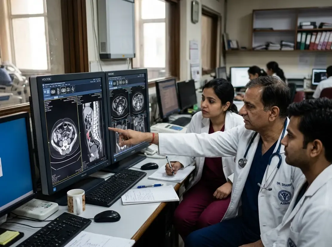
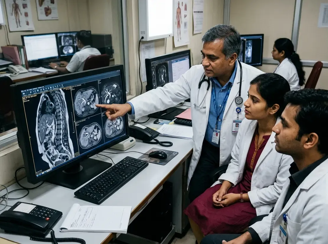
Frontotemporal dementia causes profound changes in behavior, personality, and language function. Consequently, clinicians often struggle with early frontotemporal dementia diagnosis because symptoms overlap with psychiatric illnesses and Alzheimer's disease. Recent advancements in computational neuroscience now offer promising diagnostic tools. Specifically, artificial intelligence algorithms can analyze structural and functional brain scans to detect subtle patterns of cerebral atrophy. A comprehensive systematic review and meta-analysis recently evaluated the diagnostic accuracy of these automated systems.
High Diagnostic Accuracy Across Neuroimaging Modalities
The meta-analysis examined 23 diagnostic studies that included 3,670 human subjects. Researchers pooled data from structural MRI, functional MRI, and PET scans to measure model performance against cognitively normal controls. Overall, AI models demonstrated an impressive pooled sensitivity of 91% and a pooled specificity of 92%. In addition, the summary area under the curve reached 0.96. These findings indicate that automated algorithms can reliably separate patients with dementia from healthy individuals. Furthermore, accuracy remained consistently high across all neuroimaging modalities, ranging from 0.94 to 0.98. Therefore, both structural imaging and metabolic functional scans provide viable inputs for automated pattern classification.
Frontotemporal Dementia Diagnosis Across Clinical Subtypes
Frontotemporal lobar degeneration presents through distinct clinical phenotypes. Notably, AI algorithms achieved their highest accuracy when identifying semantic variant primary progressive aphasia. For this language variant, the pooled area under the curve reached 0.98. This remarkable success reflects the distinct, asymmetrical anterior temporal lobe atrophy typical of the disorder. However, distinguishing behavioral variant frontotemporal dementia often presents greater algorithmic challenges. Patients with early behavioral changes frequently show subtle or variable structural changes on standard imaging. Thus, training algorithms to detect heterogeneous behavioral presentations remains an ongoing priority.
Current Methodological Limitations and Clinical Challenges
Despite these impressive diagnostic figures, substantial hurdles prevent immediate clinical deployment. For instance, rigorous quality appraisal using the PROBAST plus AI tool revealed widespread high risk of bias across included studies. Many investigations relied on retrospective cohorts and lacked independent external validation. Moreover, researchers observed a slight declining trend in reported accuracy from 2009 to 2025. This temporal trend likely reflects more rigorous validation standards and larger test datasets in recent years. In clinical settings, doctors must differentiate frontotemporal dementia from other dementias rather than healthy controls. Because most published models only evaluate dementia versus normal controls, their real-world differential diagnostic utility remains uncertain.
Future Directions and Practical Implementation
Clinicians should view current AI neuroimaging tools as promising diagnostic adjuncts rather than autonomous systems. Consequently, prospective multicenter trials must validate these algorithms across diverse populations and scanner platforms. In countries like India, where dementia specialist access remains limited, validated decision-support tools could substantially streamline diagnostic pathways. However, successful integration requires explainable artificial intelligence models that provide interpretable anatomical heatmaps. Ultimately, combining neuroimaging AI with clinical assessments, genetic screening, and fluid biomarkers will provide the most dependable diagnostic framework.
Frequently Asked Questions
Q1: How accurate is AI in detecting frontotemporal dementia from neuroimaging?
Meta-analysis data shows that AI models achieve a pooled sensitivity of 91% and specificity of 92%, with an area under the curve of 0.96 when distinguishing patients from cognitively normal controls.
Q2: Which subtype of frontotemporal dementia does AI identify most accurately?
AI models perform best when diagnosing semantic variant primary progressive aphasia, achieving an area under the curve of 0.98 due to distinct temporal lobe atrophy patterns.
Q3: Can clinicians currently rely on AI models as standalone diagnostic tools for dementia?
No, experts recommend using these tools only as investigational adjuncts because existing studies show high risk of validation bias and lack prospective real-world testing.
References
- Li Y et al. AI-based diagnosis of frontotemporal dementia using neuroimaging: a systematic review and meta-analysis. Eur Radiol. 2026 Oct 31. doi: 10.1007/s00330-026-13016-2. PMID: 42965691.
- Scarpina F et al. Frontotemporal dementia: a systematic review of artificial intelligence approaches in differential diagnosis. Front Aging Neurosci. 2025 Apr 9;17:1547727. doi: 10.3389/fnagi.2025.1547727.
- Lam KH et al. Use of Artificial Intelligence in Imaging Dementia. Diagnostics (Basel). 2024 Nov 27;14(23):2664. doi: 10.3390/diagnostics14232664.