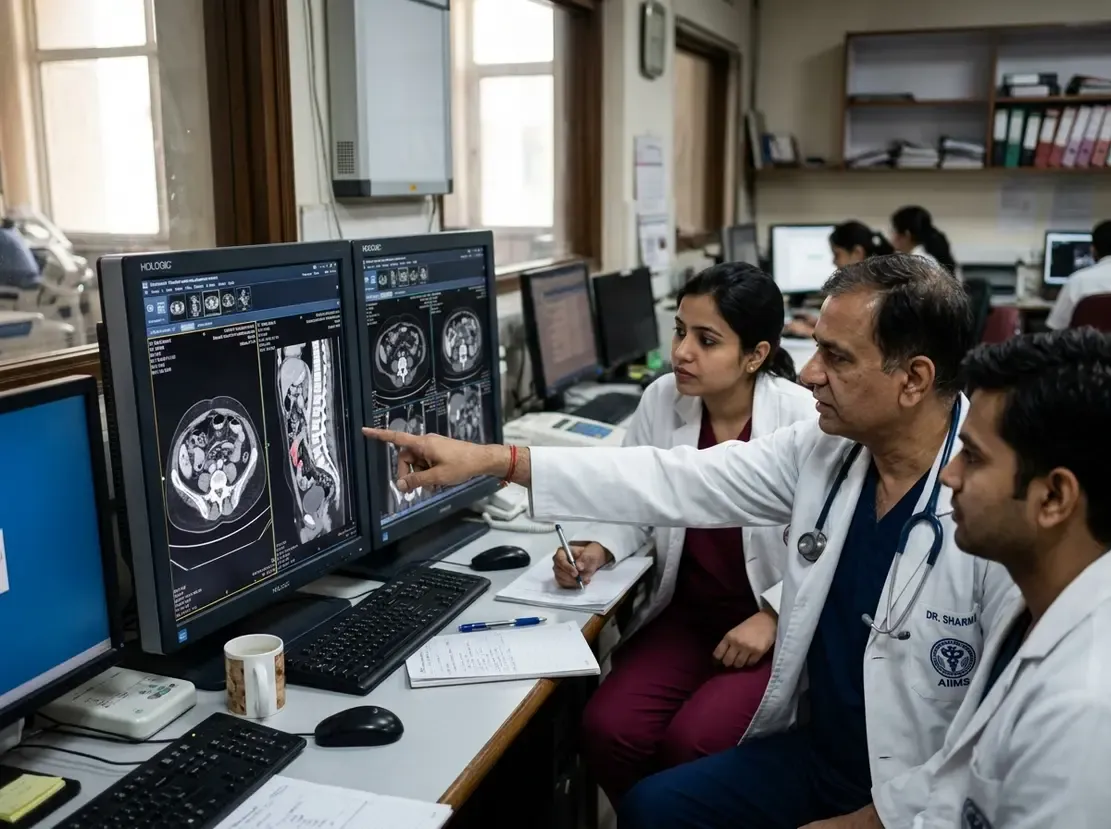
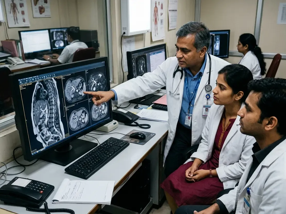
Comprehensive Guide to Hand and Wrist Vascular Imaging
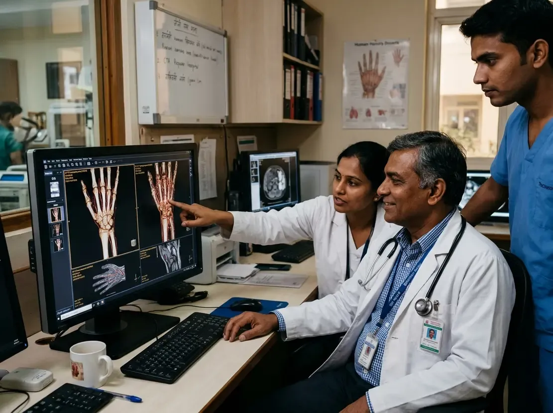
Vascular disorders of the distal upper extremity present complex diagnostic challenges for clinicians. Although rich collateral networks protect the palm, superficial vessels remain vulnerable to severe trauma. Consequently, timely evaluation prevents permanent tissue ischemia and functional limb disability. Modern hand vascular imaging offers precise roadmaps for surgical and endovascular planning. Therefore, clinicians must select appropriate diagnostic tools based on clinical urgency and vessel size.
Anatomical Nuances and Clinical Presentation
The superficial and deep palmar arches demonstrate extensive anatomical variability. For example, persistent median arteries occur in up to twenty percent of individuals. This persistent vessel can cause carpal tunnel syndrome or thrombose following local trauma. In addition, repetitive mechanical trauma frequently compromises the ulnar artery at Guyon canal. Workers using vibrating tools often develop digital vasospasm and secondary intimal hyperplasia. As a result, patients experience cold intolerance, fingertip ulceration, and localized pain.
Core Modalities in Hand Vascular Imaging
High-resolution ultrasound serves as an outstanding initial screening tool at the bedside. Clinicians utilize Doppler interrogation to detect occlusions, pseudoaneurysms, and turbulent flow patterns. Furthermore, computed tomography angiography provides rapid, high-spatial-resolution evaluation during acute traumatic emergencies. It delineates bony fractures alongside vessel disruptions without requiring prolonged acquisition times. In contrast, magnetic resonance angiography excels in characterizing soft tissue masses and slow-flow vascular malformations. Finally, digital subtraction angiography remains the reference standard for definitive therapeutic intervention.
Emerging Technologies: Photon-Counting CT and Dynamic Angiography
New imaging platforms significantly enhance our ability to visualize diminutive digital arteries. Specifically, photon-counting detector CT achieves ultra-high spatial resolution with markedly reduced beam-hardening artifacts. This advancement allows pristine visualization of tiny digital branches near metallic orthopedic hardware. Moreover, time-resolved dynamic magnetic resonance angiography captures hemodynamic flow in real time. Radiologists separate early arterial filling from venous contamination with exquisite clarity. Consequently, these advanced protocols optimize preoperative assessment before delicate composite tissue allotransplantation.
Pathology Spectrum: From Trauma to Vascular Malformations
Clinicians encounter diverse vascular pathologies ranging from hypothenar hammer syndrome to inflammatory vasculitis. For instance, blunt palmar force causes ulnar artery aneurysms and distal digital embolization. Similarly, systemic diseases like systemic sclerosis and thromboangiitis obliterans cause widespread arterial occlusions. Radiologists must also distinguish low-flow venous malformations from aggressive, high-flow arteriovenous shunts. Therefore, comprehensive multimodality imaging guides appropriate sclerotherapy or surgical resection while preventing limb loss.
Frequently Asked Questions
Q1: What makes hand vascular imaging challenging for clinicians?
Hand arteries have extremely small calibers and extensive normal anatomical variations across individuals. In addition, bone structures and patient motion frequently create imaging artifacts during diagnostic scans.
Q2: When should clinicians consider photon-counting CT for wrist pathology?
Clinicians choose photon-counting CT when evaluating microscopic digital vessels adjacent to metal implants. Furthermore, this modality provides superior spatial resolution while lowering total radiation dose.
Q3: Which vascular anomalies most commonly affect the upper extremity?
Venous malformations represent the most common low-flow vascular anomalies in the pediatric and adult hand. However, clinicians also encounter high-flow arteriovenous malformations and benign hemangiomas.
References
- Bretzman J et al. Vascular Abnormalities in the Hand and Wrist: State-of-the-Art Multimodality Imaging. Radiographics. 2026 Nov undefined. doi: 10.1148/rg.250210. PMID: 42955840.
- Ek ETM, Suh N, Carlson MG. Vascular anomalies of the hand and wrist. J Am Acad Orthop Surg. 2014 Jun;22(6):352-360. doi: 10.5435/JAAOS-22-06-352. PMID: 24860131.