Neurosurgeons often face significant challenges when trying to identify tumor boundaries during surgery. Consequently, accurate preoperative tools for glioma fluorescence prediction are essential for effective surgical planning. A recent study compared 7-Tesla magnetic resonance spectroscopic imaging (MRSI) against standard contrast-enhanced MRI and PET scans. This high-field technology offers a much deeper look into the metabolic landscape of brain tumors. Therefore, clinicians can now anticipate which areas will glow under special surgical lights.
The Role of 7-Tesla MRSI
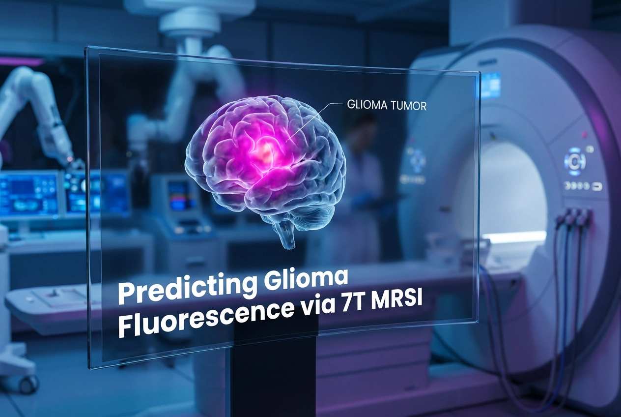
The research analyzed 43 patients with various glioma grades who underwent 7-T MRSI. Specifically, the team examined metabolic ratios to identify tumors that would show 5-ALA fluorescence. They found that 5-ALA-positive tumors exhibit distinct chemical signatures. For instance, significantly lower mI/tNAA levels often signal positive fluorescence. This ultra-high-field imaging provides higher spatial resolution than traditional scanners. Thus, it can pinpoint aggressive regions within a tumor more accurately. Doctors can use this data to target the most malignant tumor parts.
Comparing Tools for Glioma Fluorescence Prediction
How does 7-T MRSI compare to established tools like PET and standard MRI? Interestingly, MRSI outperformed contrast enhancement in predicting intraoperative fluorescence. While PET tumor-to-normal ratios (TNR) showed high sensitivity, MRSI maintained superior specificity. Therefore, surgeons might gain more clarity by using metabolic data. This is particularly valuable for non-enhancing gliomas where standard MRI results are often ambiguous. Integrating these advanced imaging markers can lead to more aggressive yet safer resections. Furthermore, it helps in selecting the right patients for 5-ALA administration.
Clinical Impact on Brain Surgery
Standard MRI often fails to show the full extent of non-enhancing gliomas. However, metabolic imaging identifies high-grade features before any physical changes occur. Thus, doctors can plan resections more effectively using these preoperative insights. Using 7-T MRSI technology might improve long-term patient outcomes. Furthermore, it reduces the need for multiple invasive diagnostic procedures. This advancement represents a significant step forward in personalized neuro-oncology. Surgeons can now achieve better results by trusting chemical biomarkers.
Frequently Asked Questions
Q1: Why is 5-ALA fluorescence important in glioma surgery?
It helps surgeons visualize tumor tissue in real-time under blue light, facilitating a more complete removal of the mass.
Q2: How does 7-Tesla MRSI differ from standard MRI?
7-Tesla MRSI offers much higher spatial resolution. It also detects specific metabolites that indicate tumor aggressiveness which are invisible on standard scanners.
Q3: Can MRSI predict fluorescence in non-enhancing tumors?
Yes, it identifies metabolic changes in non-enhancing gliomas that correlate with 5-ALA positivity, aiding the resection of difficult-to-see tumors.
References
- Huskic S et al. Preoperative prediction of 5-ALA fluorescence in gliomas: comparison of 7-Tesla magnetic resonance spectroscopic imaging, contrast-enhancement on MRI, and positron emission tomography. Eur Radiol. 2026 Mar 10. doi: 10.1007/s00330-026-12430-w. PMID: 41805721.
- Stummer W et al. Fluorescence-guided surgery with 5-aminolevulinic acid in neuro-oncology. Neurosurg Clin N Am. 2011.
- Galldiks N et al. PET imaging of gliomas. Nat Rev Clin Oncol. 2021.