Imaging Hand Vascular Abnormalities: A Diagnostic Guide
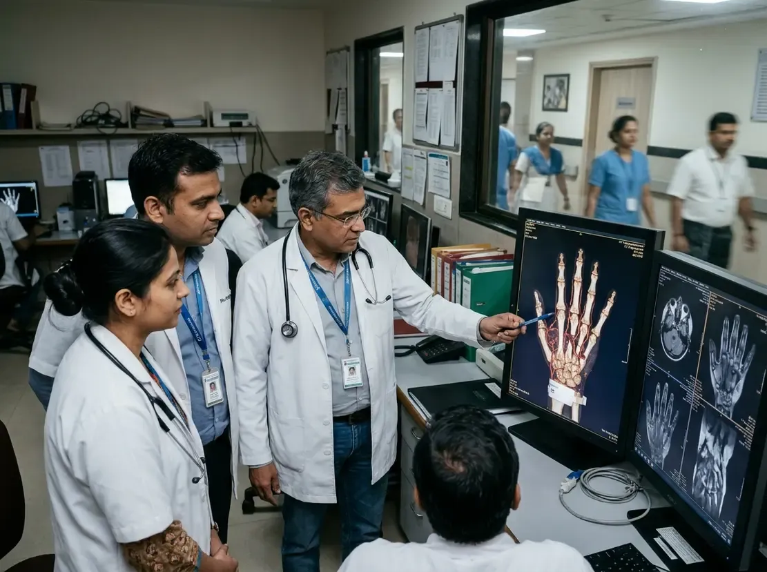
Although hand and wrist pathologies occur less frequently than lower extremity disorders, hand vascular abnormalities can cause significant functional morbidity. Therefore, early recognition remains vital to preserve hand function and avoid tissue loss. The distal upper limb possesses a rich collateral network that protects against ischemia. However, its superficial structures remain highly vulnerable to occupational trauma and repetitive impact injuries.
Anatomy and Hand Vascular Abnormalities Spectrum
First, clinicians must understand the complex arterial architecture of the hand and wrist. Anatomical variations, such as a persistent median artery, frequently alter normal perfusion patterns. Furthermore, blunt microtrauma can induce hypothenar hammer syndrome in manual workers. Similarly, vibrating tools can trigger debilitating hand-arm vibration syndrome over time. In addition, clinicians regularly encounter atherosclerotic occlusions, systemic vasculitis, embolisms, and frostbite. Vascular anomalies also represent a substantial diagnostic category, including malformations and vascular neoplasms.
Imaging Modalities for Hand Vascular Abnormalities
Consequently, multimodality imaging plays a crucial role in accurate assessment. High-resolution ultrasound serves as an excellent initial tool because it evaluates vessel patency in real time. Additionally, CT angiography provides detailed volumetric roadmaps of the delicate digital arteries. Emerging technologies like photon-counting CT further improve spatial resolution and reduce calcium blooming. Meanwhile, dynamic magnetic resonance angiography reveals flow dynamics without exposing patients to ionizing radiation. When interventional treatment is necessary, digital subtraction angiography delivers unmatched diagnostic and therapeutic capabilities. Moreover, these imaging strategies guide vascular mapping during complex hand transplantation procedures.
Frequently Asked Questions
Q1: What causes hypothenar hammer syndrome?
Repetitive blunt trauma to the hypothenar eminence injures the ulnar artery as it crosses the hook of the hamate. Consequently, this trauma leads to thrombosis, aneurysm formation, or distal digital ischemia.
Q2: Why is ultrasound a primary imaging modality for hand vascular abnormalities?
Ultrasound provides accessible, noninvasive, and real-time hemodynamic evaluation of superficial vessels. Furthermore, Doppler imaging easily identifies aneurysms, occlusions, and flow direction at the bedside.
Q3: What role does photon-counting CT play in hand vascular imaging?
Photon-counting CT offers ultra-high spatial resolution for tiny digital arteries. Therefore, it substantially reduces blooming artifacts from adjacent bones and vascular calcifications.
References
- Bretzman J et al. Vascular Abnormalities in the Hand and Wrist: State-of-the-Art Multimodality Imaging. Radiographics. 2026 Nov undefined. doi: 10.1148/rg.250210. PMID: 42955840.
- Ek ET, Suh N, Carlson MG. Vascular anomalies of the hand and wrist. J Am Acad Orthop Surg. 2014;22(6):352-360. doi:10.5435/JAAOS-22-06-352.
- Kransdorf MJ, Turner-Stepahin S, Merritt WH. Magnetic resonance angiography of the hand and wrist: Evaluation of patients with severe ischemic disease. J Reconstr Microsurg. 1998;14(2):77-81.