Radiology is at the heart of modern diagnostic medicine. Whether in emergency care, oncology, obstetrics, or neurology, timely and accurate imaging guides clinical decisions across disciplines. For practising MBBS and MD doctors, mastering the full range of imaging modalities is not merely an academic goal – it is a clinical imperative. Understanding the nuances of each technique, from when to use them to how to interpret them, is key to delivering informed, confident diagnoses.
This article breaks down the essential radiology imaging techniques that every radiologist must be adept at.
Why Imaging Modality Mastery Matters
Radiological imaging is not one-size-fits-all. Each modality serves a specific diagnostic purpose based on tissue type, clinical urgency, patient safety, and suspected pathology. Being able to select and interpret the appropriate modality is foundational to the work of every radiologist.
As imaging grows more integrated into multidisciplinary care and technology continues to evolve, the ability to work fluently across multiple imaging platforms becomes a defining skill. Doctors must not only understand the basics of imaging modalities but also learn how to integrate imaging data into real-time clinical reasoning.
The Core Types of Imaging Modalities in Radiology
1. X-Ray: The Diagnostic Entry Point
X-rays remain the most widely utilised imaging modality for the initial evaluation of skeletal trauma, chest conditions, and post-operative changes. Despite being foundational, proficiency in interpreting subtle radiographic findings and understanding principles of image acquisition is essential for accurate diagnosis.
For clinicians seeking to strengthen their radiological interpretation skills, the Certification Course in Clinical Imaging offered by OC Academy provides comprehensive training. This course covers various imaging modalities, including X-ray, and emphasises the development of diagnostic competencies through structured modules and case-based learning.
2. Ultrasound: Real-Time, Radiation-Free Insight
Ultrasound is central to imaging in obstetrics, gynaecology, hepatobiliary systems, and vascular studies. Its radiation-free nature and bedside accessibility make it especially valuable in emergency and antenatal care. Competence in probe handling and interpreting dynamic anatomy in real-time is a core radiology skill.
The Certificate Course in Gynaecology and Obstetrics Radiology offers structured training in both transabdominal and transvaginal scanning techniques, helping doctors develop applied ultrasound expertise.
3. CT Scan: The Cross-Sectional Workhorse
CT (Computed Tomography) plays a vital role in acute care settings, offering rapid, high-resolution evaluation of complex anatomical structures. It is particularly indispensable in trauma, stroke, thoracic, and abdominal imaging. Mastery of CT protocols, contrast phases, and multiplanar reconstruction techniques is essential for radiologists aiming to provide precise, timely diagnoses.
To support this skill development, OC Academy’s Certificate Course in Thoracic & Respiratory Radiology offers focused, case-based training in CT chest interpretation. The curriculum covers high-yield patterns such as interstitial lung disease (ILD), pulmonary embolism, and HRCT findings, making it ideal for doctors looking to enhance their expertise in thoracic imaging through structured, clinically relevant learning.
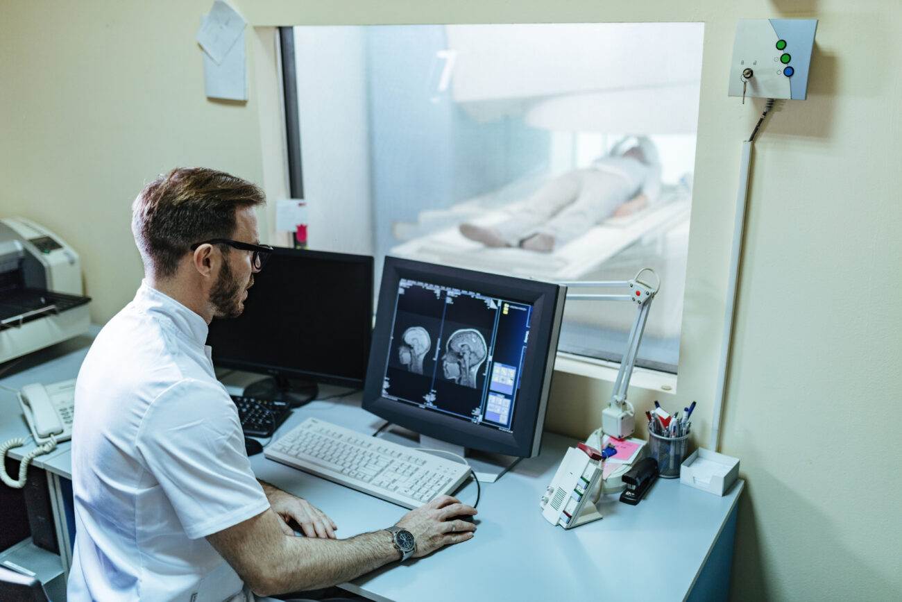
4. MRI: Precision Imaging for Soft Tissue and Neurology
Magnetic Resonance Imaging (MRI) is crucial for soft-tissue evaluation, neuroimaging, musculoskeletal disorders, and oncology follow-up. Its ability to delineate tissue contrast without ionising radiation makes it an advanced but necessary skillset.
Radiologists must learn about pulse sequences, signal intensities, and protocol planning across body systems.
5. PET and Hybrid Imaging: Functional Meets Anatomical
Positron Emission Tomography (PET), often paired with CT or MRI, delivers functional information critical to oncology, neurology, and cardiology. It aids in cancer staging, treatment response monitoring, and metabolic assessment. Proficiency requires understanding radiotracer pharmacokinetics and correlating functional data with anatomical imaging.
Though used more selectively, PET-CT and PET-MRI are increasingly integrated into advanced diagnostic workflows.
Learning Modalities in Practice: Beyond Theoretical Knowledge
Understanding the types of imaging modalities in radiology is only the starting point. True mastery lies in developing diagnostic confidence through accurate interpretation, structured reporting, and meaningful clinical correlation.
This level of competence comes not just from textbooks, but from structured, case-based learning that simulates real clinical practice. Programmes that integrate modality-specific modules with practical image analysis help doctors move from foundational knowledge to informed decision-making in varied clinical contexts.
For instance, a focused Certificate Course in Head and Neck Radiology, focused on modality selection, imaging anatomy, and diagnostic protocols, can help doctors strengthen their core interpretation skills in one of the most anatomically complex regions of the body.
Building Radiology Imaging Skills: Step-by-Step
Mastering radiology imaging skills requires more than just exposure to complex cases—it demands a structured, methodical progression. Doctors can build lasting competence by following a deliberate approach:
- Start with the basics: Develop a strong understanding of normal anatomy across all major imaging modalities.
- Correlate with clinical findings: Always interpret images in a clinical context to ensure diagnostic relevance.
- Practice structured reporting: Learn to communicate findings clearly and consistently using standardised formats.
- Review a wide range of cases: Exposure to diverse pathologies sharpens diagnostic judgement.
- Seek expert feedback: Engaging with mentor-guided training or case discussions reinforces accuracy and builds clinical confidence.
How OC Academy Helps Radiologists Upskill
For practising doctors in India aiming to enhance their diagnostic expertise, OC Academy provides structured, academically rigorous courses designed to fit alongside clinical commitments. These courses for radiology are built to support professional development without disrupting existing practice.
Key features include:
- Subspecialty-oriented modules covering thoracic imaging, MSK, neuroimaging, gynaecologic ultrasound, and more
- Flexible, self-paced learning formats are ideal for working professionals
- Expert-guided pathways aligned with international exam prep
- Certification mapped to globally recognised academic benchmarks
Whether a doctor is building core radiology competencies or exploring subspecialised domains, these courses offer a clear, practical route to continuous clinical advancement.
Conclusion
In an era where diagnostic precision is integral to clinical outcomes, mastering the full spectrum of imaging modalities is essential for every radiologist. From foundational tools like X-rays and ultrasound to advanced platforms such as CT, MRI, and PET, each modality offers unique insights that shape medical decision-making across specialities.
For MBBS and MD doctors, building radiology imaging skills is no longer optional—it’s a clinical necessity. Structured, case-based learning that bridges theoretical understanding with practical interpretation is the most effective way to achieve diagnostic confidence.
With access to flexible, academically grounded programmes, radiologists can continually sharpen their skills without interrupting clinical responsibilities. As imaging becomes increasingly central to multidisciplinary care, those equipped with both technical competence and clinical insight will lead the next generation of diagnostic medicine.
FAQs
1. What are the different types of imaging modalities in radiology, and why is it important to learn?
Radiology includes several core imaging modalities – X-ray, ultrasound, CT, MRI, and PET – each suited to specific clinical applications. Mastering them enables radiologists to select the right modality for each case, interpret findings with confidence, and contribute meaningfully to diagnosis and treatment planning. A strong foundation across modalities is essential for modern radiology practice.
2. How can doctors improve their radiology imaging skills while continuing clinical work?
Practising doctors can upskill through flexible, self-paced radiology courses that combine theory with real-world case interpretation. These programmes often focus on essential radiology imaging techniques such as CT, MRI, and ultrasound, enabling learners to acquire skills while remaining in their clinical roles.
3. Why is it necessary to understand the basics of imaging modalities before focusing on subspecialties?
Before diving into complex subspecialised areas like neuroimaging or thoracic radiology, it’s important to master the basics of each imaging technique. This includes learning normal anatomy, modality selection, and interpretation principles. A strong foundational skillset ensures more accurate reporting and better integration into multidisciplinary teams.
4. Which imaging modalities are most commonly used in day-to-day radiology practice?
In everyday practice, X-rays and ultrasound are often the first-line modalities, especially in trauma, obstetrics, and emergency care. CT is widely used in acute and cross-sectional imaging, while MRI is preferred for soft tissue, neurological, and musculoskeletal assessments. Each plays a distinct and critical role in clinical workflows.
5. How important is it for radiologists to stay updated with evolving imaging technologies?
Staying current with imaging technologies is essential for radiologists since developments such as AI implementation, hybrid imaging, and newer protocols are constantly redefining clinical practice. Regular upskilling ensures diagnostic accuracy, improves patient outcomes, and keeps radiologists aligned with global standards and modern healthcare demands.