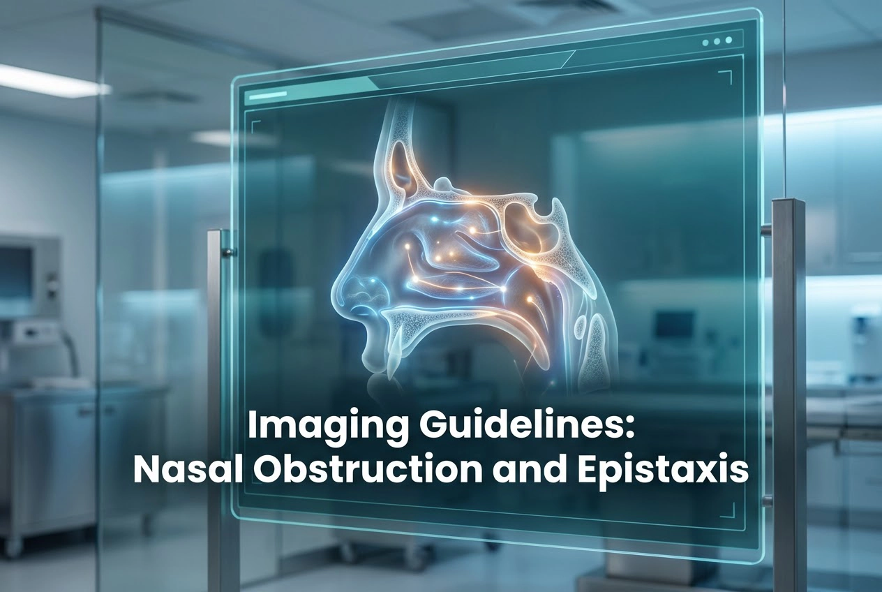
Imaging in nasal obstruction helps clinicians identify the root cause of breathing issues when initial exams are unclear. While many cases stem from common anatomical variations, some involve serious tumours or vascular disorders. Therefore, understanding the correct use of radiology is vital for every modern practitioner. This guide explores recent European Society of Head and Neck Radiology (ESHNR) recommendations to help you streamline patient care effectively.
Optimal Use of Imaging in Nasal Obstruction
Physicians should always begin with a detailed clinical history and nasal endoscopy. However, when these tools do not provide a clear answer, advanced imaging becomes necessary. Computed tomography (CT) stands as the primary modality for evaluating structural causes. Specifically, it excels at showing bony pathology and complex inflammatory diseases. Moreover, surgeons rely on CT for precise anatomical mapping before any intervention. Consequently, this leads to better surgical outcomes and fewer complications.
Magnetic resonance imaging (MRI) serves as a powerful complementary tool to CT scans. While CT captures bone details, MRI offers superior soft tissue characterization. Additionally, radiologists recommend MRI when they suspect a malignancy or intracranial extension. Thus, combining these modalities provides a comprehensive view of the sinonasal region. By selecting the right scan at the right time, you ensure that patients receive the most accurate diagnosis possible.
Advanced Imaging Protocols for Epistaxis
Doctors generally do not require routine imaging for a simple nosebleed. Nevertheless, clinicians should order scans for patients with severe, recurrent, or posterior bleeding. In these cases, imaging helps localize the bleeding source and guides interventional planning. Specifically, clinicians prefer CT angiography for vascular assessment. This high-resolution scan allows for the detection of aneurysms or tumors that cause persistent hemorrhage. Furthermore, CTA helps interventional radiologists plan life-saving embolization procedures.
Frequently Asked Questions
Q1: When should a doctor order a CT scan for nasal obstruction?
Doctors should order a CT scan when clinical history and endoscopy are inconclusive or when persistent symptoms suggest bony pathology. CT holds special importance for surgical planning because it reveals complex anatomical variations.
Q2: Is MRI better than CT for evaluating epistaxis?
Usually, clinicians prefer CT angiography for vascular assessment in severe epistaxis. However, MRI is superior for evaluating underlying soft tissue tumors that might be the primary cause of recurrent bleeding.
Q3: Should imaging be used for every case of a nosebleed?
No, most nosebleeds are minor and resolve with local treatment. Specialists strictly reserve imaging for severe, recurrent, or posterior epistaxis where clinicians suspect a vascular or neoplastic cause.
References
- Péporté ARJ et al. ESR Essentials: imaging in nasal obstruction and epistaxis-practice recommendations by the European Society of Head and Neck Radiology. Eur Radiol. 2026 Feb 16. doi: 10.1007/s00330-025-12305-6. PMID: 41697295.
- Tunkel DE et al. Clinical Practice Guideline: Nosebleed (Epistaxis). Otolaryngol Head Neck Surg. 2020.
- Gupta K et al. Computed Tomographic Evaluation of Sinonasal Variations in the Indian Population. Indian J Otolaryngol Head Neck Surg. 2024.