Can MRI Detect Painful Post-THA Abductor Tendon Tears?
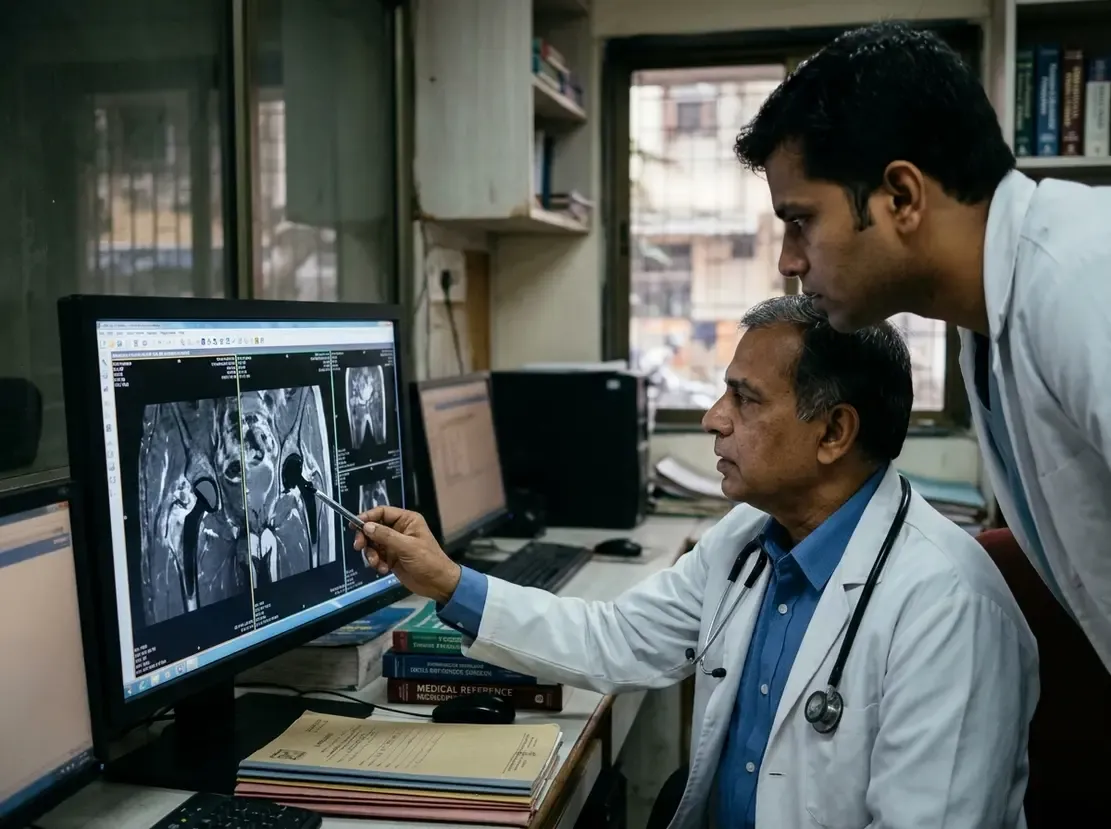
Persistent trochanteric pain and gait instability often trouble patients following total hip arthroplasty. Specifically, lateral surgical approaches frequently compromise the gluteus medius and gluteus minimus tendons. Consequently, detecting hip abductor tendon tears remains essential for guiding revision surgery. Advanced magnetic resonance imaging offers exceptional anatomical detail despite adjacent metallic implants. A landmark study now evaluates how effectively MRI detects these debilitating tendon lesions.
Diagnostic Precision for Hip Abductor Tendon Tears
The retrospective investigation examined 36 patients undergoing revision surgery after primary arthroplasty. Two experienced musculoskeletal radiologists independently evaluated preoperative MRI scans. Intraoperative inspection served as the definitive reference standard for tendon integrity.
Notably, Reader 1 demonstrated diagnostic sensitivities ranging from 0.86 to 1.00. Furthermore, Reader 1 attained specificities between 0.93 and 1.00, alongside accuracies reaching 0.92 to 0.97. Similarly, Reader 2 achieved sensitivities between 0.86 and 1.00 with specificities up to 1.00. Consequently, both readers proved the high reliability of magnetic resonance imaging in clinical practice.
Key Imaging Features and Clinical Correlations
Surgeons often struggle to differentiate true structural tears from benign postoperative scarring. However, regression models revealed definitive imaging biomarkers linked to surgical failure. Specifically, discrete tendon defects strongly predicted both partial- and full-thickness tears. Gluteus medius tears correlated with an odds ratio of 35.5. In addition, gluteus minimus disruptions demonstrated an odds ratio of 30.0.
Moreover, peritrochanteric fluid collections and severe fatty muscle degeneration provided crucial diagnostic clues. Radiologists routinely grade abductor fatty atrophy using the established Goutallier classification system. Therefore, integrating qualitative tendon architecture with muscle degeneration improves preoperative planning. These findings allow orthopedic teams to select appropriate candidates for surgical abductor repair.
Frequently Asked Questions
Q1: Why does a lateral surgical approach increase the risk of abductor tears?
The transgluteal approach splits or detaches the gluteus medius and minimus tendons during joint exposure. Consequently, repair failure or denervation can cause persistent tendon insufficiency and trochanteric pain.
Q2: How does MRI overcome metal artifact around hip prostheses?
Modern protocols apply optimized metal artifact reduction sequences. Therefore, radiologists can clearly visualize soft tissues and peritrochanteric fluid near metallic components.
Q3: What imaging sign best predicts a confirmed abductor tendon tear?
Discrete tendon discontinuity or defect exhibits the strongest association with surgically confirmed tears. Furthermore, marked peritrochanteric fluid and muscle fatty atrophy corroborate the diagnosis.
References
- Marth AA et al. MRI for the detection of hip abductor tendon tears in symptomatic patients with total hip arthroplasty following a lateral surgical approach. Eur Radiol. 2026 Oct 30. doi: 10.1007/s00330-026-12856-2. PMID: 42961652.
- Pfirrmann CW et al. Abductor tendons and muscles assessed at MR imaging after total hip arthroplasty in asymptomatic and symptomatic patients. Radiology. 2005 Jun;235(3):969-76.
- Cvitanic O et al. MRI diagnosis of tears of the hip abductor tendons (gluteus medius and gluteus minimus). AJR Am J Roentgenol. 2004 Jan;182(1):137-43.