Muscle MRI and AI: Refining Distal Myopathy Diagnosis
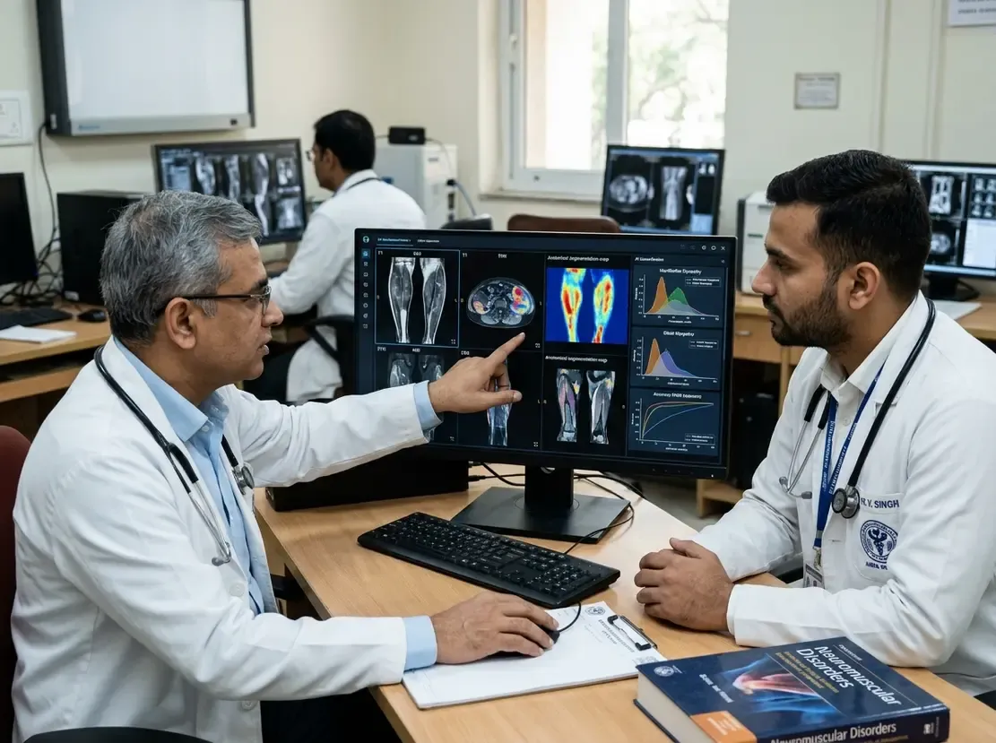
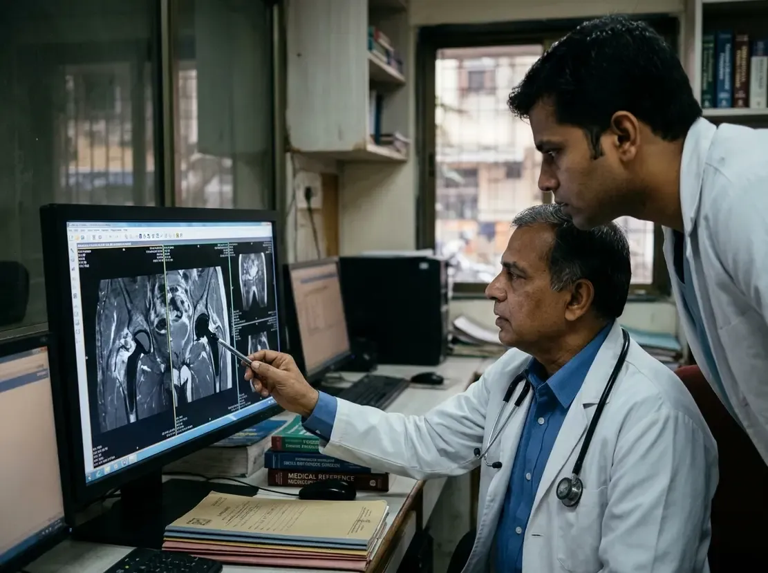
Distal and myofibrillar myopathies present a formidable diagnostic challenge because symptoms overlap substantially across genetic subtypes. Fortunately, identifying specific muscle MRI patterns helps clinicians distinguish these complex hereditary muscle disorders with remarkable clarity. A multicentre study evaluated 165 patients across 19 different disease genes. Overall, investigators systematically reviewed 6,350 skeletal muscles. Consequently, the authors established reproducible imaging signatures for multiple myopathy subtypes.
Key Muscle MRI Patterns in Distal and Myofibrillar Myopathies
Each genetic myopathy revealed distinctive anatomical signatures upon cross-sectional imaging. For instance, GNE myopathy showed early selective fatty replacement of the biceps femoris short head. In contrast, patients with desminopathy exhibited early involvement of the flexor hallucis longus muscle. Furthermore, scans from individuals with myotilinopathy displayed discrete intramuscular fat pockets. Importantly, these imaging signatures showed sensitivity and specificity values ranging between 75% and 100%. Therefore, qualitative MRI findings deliver dependable biomarkers for routine clinical differential diagnosis.
Integrating Machine Learning for Genetic Classification
Genetic heterogeneity often complicates traditional diagnostic workflows in neuromuscular clinics. To address this challenge, researchers trained supervised machine learning algorithms using quantitative muscle scores. Among the tested models, XGBoost demonstrated the highest overall performance. Specifically, the XGBoost algorithm correctly distinguished among 11 different genetic conditions. Moreover, the model achieved a balanced accuracy of 69% across complex diagnostic categories. Thus, artificial intelligence effectively translates subtle imaging features into reliable clinical predictions.
Clinical Implications for Practice
Diagnosing inherited myopathies in clinical practice frequently requires costly whole-exome sequencing. However, accessible imaging protocols offer rapid, non-invasive diagnostic guidance. By integrating machine learning classifiers with muscle scans, clinicians can prioritise likely genetic variants. Additionally, this combined strategy reduces reliance on invasive muscle biopsies. Consequently, neurologists can accelerate definitive diagnosis and initiate targeted supportive therapies sooner.
Frequently Asked Questions
Q1: What are the main muscle MRI patterns seen in distal myopathies?
Key patterns include early biceps femoris short head involvement in GNE myopathy and early flexor hallucis longus changes in desminopathy. Additionally, myotilinopathy typically features distinctive intramuscular fat pockets.
Q2: How does machine learning improve distal myopathy diagnosis?
Supervised machine learning models such as XGBoost analyse complex muscle involvement scores across multiple muscle groups. Consequently, these algorithms achieve high balanced accuracy and help clinicians prioritise specific genetic tests.
Q3: Can muscle MRI replace genetic testing in inherited myopathies?
No, muscle MRI does not replace confirmatory genetic testing. However, it directs focused genetic panels, minimises inconclusive findings, and avoids invasive diagnostic procedures.
References
- Bortolani S et al. Diagnostic accuracy of MRI-derived imaging biomarkers and machine learning-based classification in distal and myofibrillar myopathies. Eur Radiol. 2026 Oct 30. doi: 10.1007/s00330-026-13014-4. PMID: 42961653.
- Diaz-Manera J, Llauger J, Gallardo E, Illa I. Muscle MRI in distal myopathies: clinical applications and diagnostic algorithms. Front Neurol. 2018;9:452.
- Morrow JM, Sinclair CD, Fischmann A, et al. Muscle MRI in neuromuscular diseases: an update. Pract Neurol. 2020;20(5):370-380.