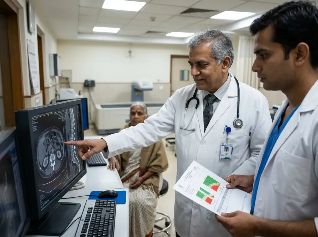
Enhancing O-RADS MRI Accuracy with Intensity Curves

Improving O-RADS MRI Diagnostic Accuracy
Accurate preoperative risk stratification of adnexal masses remains essential for guiding surgical triage. The standard O-RADS MRI framework effectively differentiates benign from malignant ovarian lesions. However, standard visual assessment of solid tissue enhancement can sometimes yield false-positive results. Consequently, investigators evaluated whether a semi-quantitative intensity curve (SIC) could improve diagnostic specificity.
A recent multicenter retrospective study analyzed 341 adnexal lesions across 321 women. Notably, solid tissue components were present in over 96% of these masses. Radiologists assigned an O-RADS MRI score using both conventional visual assessment and manual SIC analysis. As a result, researchers directly compared the clinical impact of both diagnostic methods on patient management.
Visual Assessment Versus Semi-Quantitative Intensity Curves
Standard visual assessment achieved a sensitivity of 95% and a specificity of 84% through detailed clinical imaging evaluations. Furthermore, the positive predictive value reached 77% under standard visual evaluation. In contrast, incorporating SIC increased the specificity significantly to 92%. Additionally, the positive predictive value rose to 90% when using the semi-quantitative curve method.
Therefore, semi-quantitative curve integration markedly reduced the misclassification of benign lesions. The proportion of benign masses triaged for extensive oncologic resection dropped from 16% to 9%. Moreover, readers demonstrated strong inter-reader and intra-reader agreement across both assessment modalities.
Clinical Relevance for O-RADS MRI in Practice
Differentiating complex benign masses from invasive cancers helps surgeons select the most appropriate intervention. General gynecologists can safely manage benign lesions using fertility-sparing or minimally invasive techniques. Conversely, specialized gynecologic oncologists must manage borderline and malignant neoplasms to ensure optimal outcomes, which is a key focus within advanced clinical oncology training.
Thus, integrating semi-quantitative intensity curves into routine MRI interpretation refines surgical triage. Radiologists can easily implement this manual approach without complex software tools. Ultimately, this enhanced diagnostic workflow prevents unnecessary radical surgery while maintaining excellent detection rates for malignant tumors.
Frequently Asked Questions
Q1: How does SIC improve O-RADS MRI performance?
SIC provides an objective measurement of tissue enhancement over time. Consequently, it significantly increases diagnostic specificity and positive predictive value compared to visual inspection alone.
Q2: Does using SIC compromise malignancy detection rates?
No, the protocol maintains a high sensitivity of 95%. Therefore, clinicians can confidently identify malignant lesions while reducing false-positive diagnoses.
Q3: How does this method affect surgical decision-making?
Incorporating SIC reduces unnecessary gynecologic oncology referrals for benign lesions. As a result, more patients receive appropriate, organ-sparing surgical management.
References
- Salinas-Miranda E et al. Improving diagnostic performance of O-RADS MRI: semi-quantitative intensity curves vs visual assessment of enhancement. Eur Radiol. 2026 Aug 30. doi: 10.1007/s00330-026-12742-x. PMID: 42668304.
- Sadowski EA, Thomassin-Naggara I, Rockall A, et al. O-RADS MRI Risk Stratification System: Guide for Assessing Adnexal Lesions from the ACR O-RADS Committee. Radiology. 2022;303(1):35-47.
- Thomassin-Naggara I, Poncelet E, Jalaguier-Coudray A, et al. O-RADS MRI Classification of Indeterminate Adnexal Lesions: Time-Intensity Curve Analysis Is Better Than Visual Assessment. Radiology. 2020;296(2):413-421.