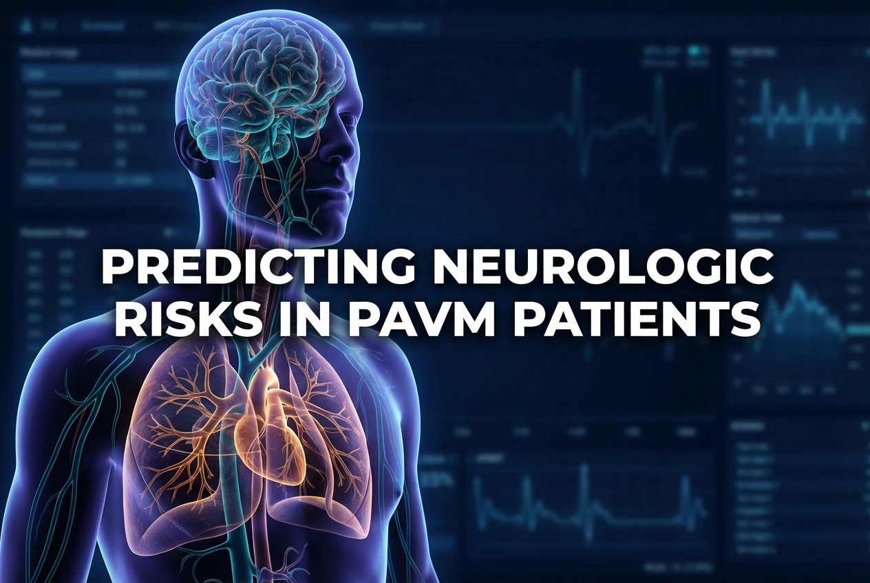
Pulmonary arteriovenous malformations (PAVMs) are abnormal connections in the lungs that create a right-to-left shunt, bypassing the pulmonary capillary filter. Consequently, this can lead to severe, life-threatening conditions. The most concerning of these are symptomatic PAVM neurologic complications, such as ischemic stroke or brain abscess. Therefore, identifying patients at high risk for these outcomes is critically important for management and timely intervention.
A recent multicenter CT-based analysis of 207 patients focused on identifying anatomic predictors of symptomatic complications in solitary, untreated PAVMs. The study compared 36 patients who had experienced a complication (neurologic group) with 171 controls. Importantly, the researchers excluded transient ischemic attacks to focus on definitive, symptomatic events.
Anatomic Predictors of PAVM Neurologic Complications
Analysis showed significant differences in certain imaging features between the two groups. Specifically, the neurologic group presented with significantly larger feeding artery diameters. The median diameter was 3.8 mm, compared to 3.0 mm in the control group. Furthermore, the sac size was also markedly larger in patients with complications, with a median of 9.9 mm versus 6.5 mm. These findings suggest that the absolute size of the AVM components significantly influences the risk profile.
Moreover, the sac-to-feeding artery (SF) ratio was another strong predictor. The median SF ratio was significantly lower in the neurologic group (1.9 vs. 2.3). This suggests that a relatively smaller sac compared to the feeding artery may increase the flow velocity or turbulent stress, thus increasing the likelihood of complications. However, no significant differences were observed in factors like patient age, sex, PAVM location, type, or the presence of intrasaccular thrombus.
Clinical Context and Management Implications
The risk of neurologic sequelae, including stroke and cerebral abscess, is well-established in PAVM patients due to paradoxical emboli—clots or bacteria bypassing the lung’s filtering action. Consequently, interventional cardiologists frequently use embolisation to block these abnormal vessels, which demonstrably reduces these severe risks. In India, numerous specialist centres offer percutaneous embolisation, which is considered a safe and effective treatment method. Therefore, determining a strict size-based threshold for intervention is essential, especially when patients are otherwise asymptomatic. The study strongly supports current clinical guidelines that often target PAVMs with a feeding artery diameter greater than 3 mm for treatment, as this size correlates with a substantially higher risk of cerebral events. This diagnostic method provides clinicians with an objective metric for risk stratification and treatment planning. Specifically, the findings reinforce the critical role of high-resolution CT in the initial diagnosis and follow-up of these patients.
Frequently Asked Questions
Q1: What are the most serious PAVM neurologic complications?
The most serious symptomatic neurologic complications identified in the study are ischemic stroke and brain abscess. These result from paradoxical emboli (clots or bacteria) traversing the PAVM and entering the systemic circulation, often reaching the brain.
Q2: What CT measurements predict a higher risk of complications?
The key anatomic predictors identified by CT are a larger feeding artery diameter (median 3.8 mm in the high-risk group), a larger sac size (median 9.9 mm), and a lower sac-to-feeding artery (SF) ratio.
Q3: How is the risk of stroke reduced in patients with a high-risk PAVM?
The primary method for reducing the risk of stroke and other complications is pulmonary AVM embolisation. This minimally invasive procedure blocks the abnormal vessel using coils or plugs, thereby restoring normal blood filtration and preventing paradoxical emboli.
References
- Shimohira M et al. Anatomic predictors of neurologic complications in solitary pulmonary arteriovenous malformations: a multicenter CT-based analysis. Eur Radiol. 2025 Dec 17. doi: 10.1007/s00330-025-12213-9. PMID: 41405690.
- Hsu YJ et al. Risk factors for cerebral complications in patients with pulmonary arteriovenous malformations: A multicenter retrospective cohort study. PLOS One. 2022 Dec 1. doi: 10.1371/journal.pone.0278457. PMID: 36454790.
- Shankar A and Singh S. Pulmonary Arteriovenous Malformation. StatPearls [Internet]. Treasure Island (FL): StatPearls Publishing; 2024.