Accurate staging of primary lung cancer is crucial for optimizing therapeutic strategies. However, identifying lymph node metastasis (LNM) remains a major clinical challenge. For this reason, researchers developed a novel PET/CT nomogram combining clinical characteristics with imaging findings. This powerful tool helps predict the risk of LNM on an individual basis.
Non-small cell lung cancer (NSCLC) staging profoundly impacts patient prognosis and treatment planning. Although advanced imaging techniques like PET/CT enhance diagnostic accuracy, a certain percentage of occult LNM still occurs. Therefore, clinicians need a more precise, non-invasive method for preoperative assessment. This is especially true for early-stage patients who might otherwise undergo unnecessary surgery or, conversely, miss vital adjuvant therapy. Importantly, using predictive models can improve the selection of candidates for minimally invasive procedures.
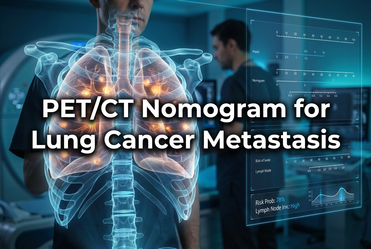
Key Features of the PET/CT Nomogram
Researchers retrospectively analyzed patient data to develop five distinct predictive models. These models all aimed to predict LNM using various combinations of data: clinical, CT, and PET findings. The key is the ability to integrate diverse information into one scoring system. For instance, common predictive factors in these nomograms include age, tumor location (central vs. peripheral), and tumor histology (e.g., adenocarcinoma). Also, the maximum standardized uptake value (SUVmax) from both the primary tumor and the suspicious lymph node are critical inputs. Significantly, studies show that combining these clinical and metabolic factors provides superior discrimination compared to using imaging or clinical factors alone.
The ultimate goal of the PET/CT nomogram is to stratify patient risk effectively. Using the nomogram, physicians gain confidence in identifying patients with a low probability of LNM who may safely proceed with limited resection or avoid invasive nodal procedures. Conversely, patients with a high predicted risk benefit from more aggressive nodal staging or upfront systemic therapy. Moreover, these personalized tools improve clinical decision-making by offering a quantitative, evidence-based score rather than relying solely on subjective criteria. Consequently, nomograms have demonstrated robust discrimination and calibration, showing excellent agreement between predicted and actual LNM outcomes.
Frequently Asked Questions
Q1: What is the gold standard used to diagnose lymph node metastasis (LNM) in this study?
The gold standard for diagnosing LNM was the cytological confirmation of samples obtained through endobronchial ultrasound-guided transbronchial needle aspiration (EBUS-TBNA).
Q2: Which factors are typically included in a PET/CT nomogram for lung cancer?
Nomograms frequently include clinical characteristics like age, tumor histology, and location. Furthermore, they incorporate imaging metrics such as the primary tumor’s maximum standardized uptake value (SUVmax) and the lymph node SUVmax.
References
- Han X et al. Development of PET/CT-clinical nomograms for predicting lymph node metastasis in primary lung cancer. Eur Radiol. 2025 Dec 17. doi: 10.1007/s00330-025-12166-z. PMID: 41405691.
- Wei X et al. A nomogram for predicting the risk of lymph node metastasis in T1–2 non-small-cell lung cancer based on PET/CT and clinical characteristics. Transl Lung Cancer Res. 2020 Apr;9(2):127-137.
- Huang K et al. 18F-FDG PET/CT radiomics nomogram for predicting occult lymph node metastasis of non-small cell lung cancer. Front Oncol. 2022 Jul 1;12:938381.