Acute gastrointestinal (GI) bleeding is a common and potentially life-threatening emergency. Consequently, rapid and accurate diagnosis is essential for effective patient management. A new study demonstrates that a Dual-Energy CT (DECT) protocol is diagnostically non-inferior to conventional triphasic CT for DECT GI bleeding assessment. This modern approach incorporates Virtual Non-Contrast (VNC) images and iodine maps instead of a true non-contrast phase. Furthermore, researchers found that this technique reduces both reading time and improves diagnostic confidence.
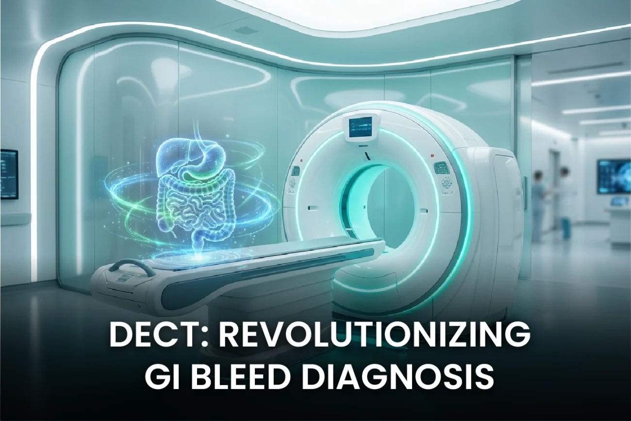
DECT GI Bleeding Protocol Outperforms Conventional CT
Conventional CT angiography (CTA) for GI bleeding typically requires a three-phase protocol: non-contrast, arterial, and portal venous. The non-contrast phase helps radiologists identify pre-existing hyperdensities, such as clotted blood, foreign bodies, or ingested material, distinguishing them from active contrast extravasation. However, obtaining this extra phase increases the patient’s radiation exposure. Virtual Non-Contrast (VNC) imaging, a powerful post-processing application of DECT, creates a synthetic image by subtracting iodine content from the contrast-enhanced data.
The study, which included 100 patients with and without GI bleeding, compared the diagnostic accuracy of the two methods. The results show compelling evidence for the DECT protocol. With conventional triphasic CT, sensitivity was 91.6% and specificity was 94.4%. Conversely, the DECT protocol achieved a sensitivity of 94.4% and a specificity of 96.0%. This confirms non-inferiority with a margin of 3%. Therefore, doctors can use the DECT method without compromising diagnostic precision. Moreover, DECT offers a significant radiation dose reduction, often cited as approximately 30%, because it eliminates the initial non-contrast scan.
Improved Efficiency and Diagnostic Confidence
Beyond maintaining accuracy, the DECT protocol introduces substantial clinical efficiency improvements. Radiologists’ diagnostic confidence saw a statistically significant increase, moving from a median score of 4 to 5 (on a 5-point scale). Likewise, the median reading time decreased from 252 seconds to 226 seconds (p < 0.001). This speed is crucial in the acute setting of GI hemorrhage. Consequently, faster diagnosis can lead to more rapid and appropriate treatment intervention.
The specialized images generated by DECT enhance the radiologist’s ability to localize the source of bleeding. VNC images effectively differentiate active bleeding (which disappears on VNC) from high-density enteric contents or hematomas (which persist on VNC). Furthermore, iodine maps and low-keV Virtual Monoenergetic Images (VMI) increase the conspicuity of subtle contrast extravasation, thereby increasing diagnostic confidence. These advanced post-processing tools solve problems often faced with conventional single-energy CT and make DECT highly useful in imaging diagnosis.
Frequently Asked Questions
Q1: What advantage does DECT offer over conventional CT for GI bleeding?
The primary advantage of DECT with the Virtual Non-Contrast (VNC) protocol is significant radiation dose reduction by eliminating the initial true non-contrast phase. Importantly, the study confirms this is achieved without sacrificing diagnostic accuracy, and it also improves diagnostic confidence and reduces reading time.
Q2: What is the primary function of Virtual Non-Contrast (VNC) images in this DECT protocol?
VNC images are critical for problem-solving. They help radiologists differentiate between active contrast extravasation (iodine), which is subtracted and therefore disappears on the VNC image, and non-iodine-containing materials like clotted blood, surgical clips, or ingested pills, which remain hyperdense on the VNC image.
References
- Oberparleiter M et al. Replacing true non-contrast imaging with DECT in GI bleeding demonstrates non-inferior diagnostic performance, reading time and confidence. Eur Radiol. 2025 Dec 17. doi: 10.1007/s00330-025-12191-y. PMID: 41405692.
- Dual-energy CT for gastrointestinal bleeding. Emerg Radiol. 2021 Apr. doi: 10.1007/s10140-021-01931-4.
- Dual-Energy CT Evaluation of Gastrointestinal Bleeding. Radiographics. 2023 May-Jun. doi: 10.1148/rg.220192.
- CT angiography for acute gastrointestinal bleeding: what the radiologist needs to know. Abdom Radiol (NY). 2022 Nov. doi: 10.1007/s00261-022-03612-z.