Can Quantitative MRI Replace Renal Mass Biopsy in RCC?
Accurate preoperative grading of renal cell carcinoma remains a critical challenge for urologists and oncologists. Fortunately, quantitative MRI in RCC has emerged as a promising non-invasive tool to predict tumor aggressiveness. Pathologists utilize the World Health Organization and International Society of Urological Pathology (WHO/ISUP) grading system to determine prognosis. However, percutaneous needle core biopsy carries minor procedural risks and suffers from sampling error. Consequently, clinicians seek reliable imaging biomarkers to distinguish indolent low-grade tumors from aggressive high-grade malignancies.
Diagnostic Accuracy of Quantitative MRI in RCC
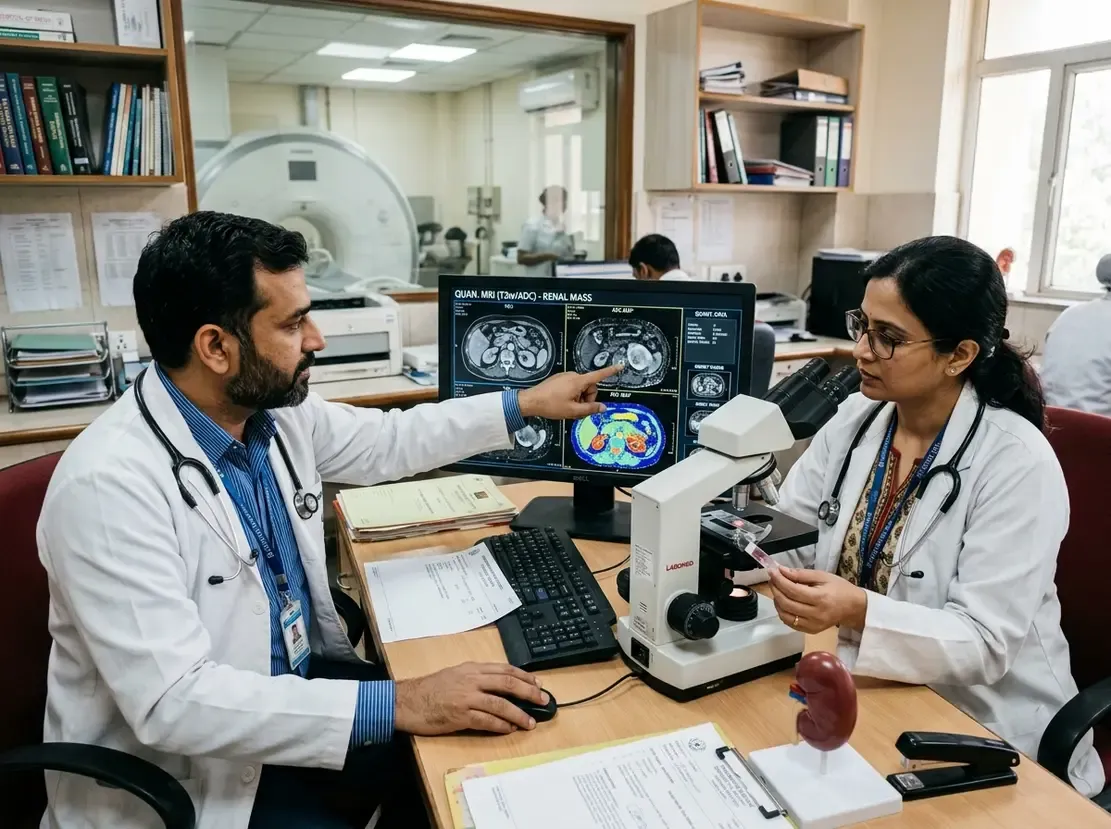
A comprehensive meta-analysis evaluated multiple quantitative imaging techniques across 20 clinical studies. In particular, researchers assessed diffusion-weighted imaging (DWI) and apparent diffusion coefficient (ADC) metrics. The pooled analysis of seven DWI studies demonstrated a sensitivity of 0.84 for detecting high-grade tumors. However, the pooled specificity reached only 0.57, yielding a summary area under the curve (AUC) of 0.71. Furthermore, low-grade tumors exhibited significantly higher ADC values than high-grade lesions, with a mean difference of 0.21 × 10⁻³ mm²/s. Therefore, while ADC maps show biological differences in cellularity, standard diffusion metrics provide only modest diagnostic accuracy when used alone.
Diffusion Imaging Versus Radiomics and Deep Learning
Advanced computational techniques appear to enhance non-invasive classification beyond conventional diffusion metrics. Specifically, five studies evaluated MRI-inclusive radiomics and deep learning models for WHO/ISUP grading. These artificial intelligence algorithms achieved a pooled sensitivity of 0.79 and a pooled specificity of 0.86. Moreover, the summary AUC reached an impressive 0.90, surpassing conventional apparent diffusion coefficient measurements. Nevertheless, significant heterogeneity exists among these algorithmic pipelines. Investigators must validate these computational tools across multi-vendor MRI scanners before adopting them in routine clinical care.
Clinical Implications for Biopsy Triage and Practice
These diagnostic findings carry direct clinical value for surgical planning and surveillance strategies. For example, clinicians can use quantitative metrics to triage patients who require confirmatory histological biopsies. Additionally, high apparent diffusion values can reassure clinicians when managing elderly patients with active surveillance. In contrast, low ADC values signal densely cellular, aggressive tissue that may warrant prompt partial or radical nephrectomy. As a result, non-invasive imaging bridges the gap between clinical risk assessment and invasive interventions. Thus, standardizing multiparametric MRI protocols across healthcare institutions remains the next crucial milestone.
Frequently Asked Questions
Q1: What is the main clinical advantage of quantitative MRI in renal cell carcinoma?
Quantitative MRI provides non-invasive functional insights into tumor cellularity. Consequently, it helps clinicians differentiate high-grade from low-grade renal cell carcinomas without immediate biopsy.
Q2: How accurate is the apparent diffusion coefficient for WHO/ISUP grading?
Standard ADC metrics demonstrate modest diagnostic performance. Specifically, meta-analytic data show a pooled sensitivity of 84% but a modest specificity of 57%, producing an AUC of 0.71.
Q3: Do radiomics and deep learning models outperform conventional diffusion MRI?
Yes, MRI-inclusive machine learning models achieve a higher diagnostic performance, with a pooled AUC of 0.90. However, substantial heterogeneity across studies requires external prospective validation.
References
- Kiani I et al. Quantitative MRI for World Health Organisation/International Society of Urological Pathology Grading of Renal Cell Carcinoma: a systematic review and diagnostic meta-analysis. Eur Radiol. 2026 Sep 18. doi: 10.1007/s00330-026-12837-5. PMID: 42758284.
- Baytok A, Ecer G, Balasar M, Koplay M. Computed tomography and magnetic resonance imaging characteristics of renal cell carcinoma: Differences between subtypes and clinical evaluation. J Clin Imaging Sci. 2025;15:10. doi: 10.25259/JCIS_160_2024.
- Pietersen PI, Lynggård Bo Madsen J, Asmussen J, et al. Multiparametric magnetic resonance imaging for characterizing renal tumors: A validation study of the algorithm presented by Cornelis et al. J Clin Imaging Sci. 2023;13:7. doi: 10.25259/JCIS_147_2022.