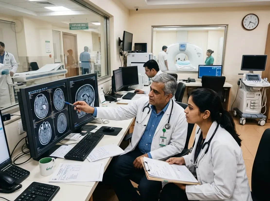
How Photon-Counting CT Is Transforming Head & Neck Scans
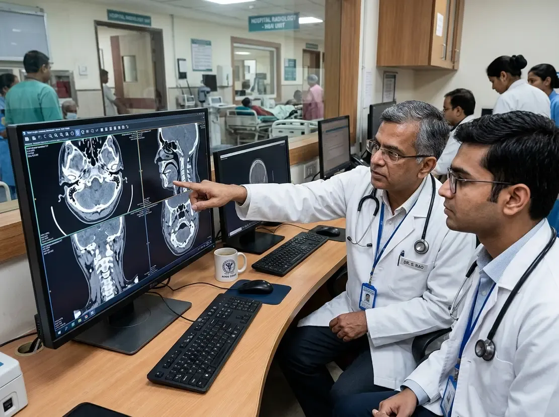
Photon-counting CT marks a transformative leap forward in head and neck imaging. Conventional energy-integrating computed tomography often struggles with spatial resolution and artifact management. In contrast, photon-counting detectors convert X-ray photons directly into electrical signals. Consequently, clinicians gain sharper image quality with substantially fewer artifacts.
Core Technical Advances of Photon-Counting CT
This imaging revolution rests on five fundamental technical pillars. First, the technology provides ultra-high spatial resolution. As a result, radiologists can delineate minute anatomic structures with remarkable clarity. Second, energy-resolving detectors eliminate electronic noise and mitigate metal artifacts. Therefore, radiologists can evaluate complex post-surgical cases more confidently. Third, the system dramatically lowers radiation exposure. Furthermore, it reduces the required volume of iodinated contrast agent. Finally, multi-energy spectral capabilities enable advanced material decomposition. For example, iodine maps and virtual non-contrast reconstructions allow precise characterization of soft tissues. Together, these five technical strengths deliver superior diagnostic accuracy.
Clinical Impact on Head and Neck Assessment
Clinicians encounter unique anatomical challenges in the head and neck region. However, superior spatial resolution significantly improves the visualization of delicate bones, including middle ear ossicles. Surgeons can also assess fine vascular branches and laryngeal cartilages with higher fidelity. In addition, artifact reduction resolves longstanding diagnostic dilemmas caused by dental hardware and surgical implants. Dentists and head-neck oncologists can examine the adjacent mucosa without obscuring scatter. Moreover, reduced radiation doses protect pediatric patients and vulnerable thyroid tissues. Similarly, minimizing contrast media volume shields patients with borderline renal function from contrast-induced nephropathy. Radiologists also leverage spectral imaging to assess carotid plaques and distinguish tumor margins from adjacent inflammatory changes. Ultimately, these clinical applications enhance surgical planning and post-treatment surveillance through specialized training available in a Certification Course In Head & Neck Radiology.
Future Prospects in Diagnostic Practice
Clinical adoption of this advanced detector platform continues to accelerate worldwide. Consequently, diagnostic protocols will increasingly incorporate routine spectral datasets without increasing scan acquisition time. Furthermore, combining photon-counting detectors with artificial intelligence reconstruction models will elevate diagnostic confidence even further. In summary, this imaging modality sets a safer and more precise benchmark for complex head and neck imaging.
Frequently Asked Questions
Q1: How does photon-counting CT reduce dental metal artifacts?
Photon-counting detectors measure individual photon energy bins directly. Consequently, specialized reconstruction algorithms filter out low-energy photon starvation, which drastically minimizes beam hardening and streak artifacts from metallic hardware.
Q2: Why is photon-counting CT particularly beneficial for pediatric head and neck imaging?
The technology offers higher dose efficiency and eliminates electronic noise. Therefore, clinicians can capture diagnostic-quality scans at substantially lower radiation doses, which significantly lowers long-term oncologic risks in children.
Q3: How does spectral imaging assist in head and neck cancer evaluation?
Spectral maps, such as virtual non-contrast and iodine concentration reconstructions, improve soft tissue differentiation. As a result, radiologists can accurately delineate tumor margins, detect invasion into cartilage, and evaluate lymph node metastases.
References
- Martín-Noguerol T et al. Photon-counting CT in head and neck radiology: bridging detector physics and clinical applications. Eur Radiol. 2026 Sep 17. doi: 10.1007/s00330-026-12910-z. PMID: 42752922.
- Benson JC, Campeau NG, Diehn FE, et al. Photon-Counting CT in the Head and Neck: Current Applications and Future Prospects. AJNR Am J Neuroradiol. 2024;45(8):1024-1033. doi: 10.3174/ajnr.A8265.
- McCollough CH, Rajendran K, Leng S, et al. The technical development of photon-counting detector CT. Eur Radiol. 2023;33(8):5321-5330. doi: 10.1007/s00330-023-09545-9.