Mastering Susceptibility-Weighted Imaging: ESR Guidelines
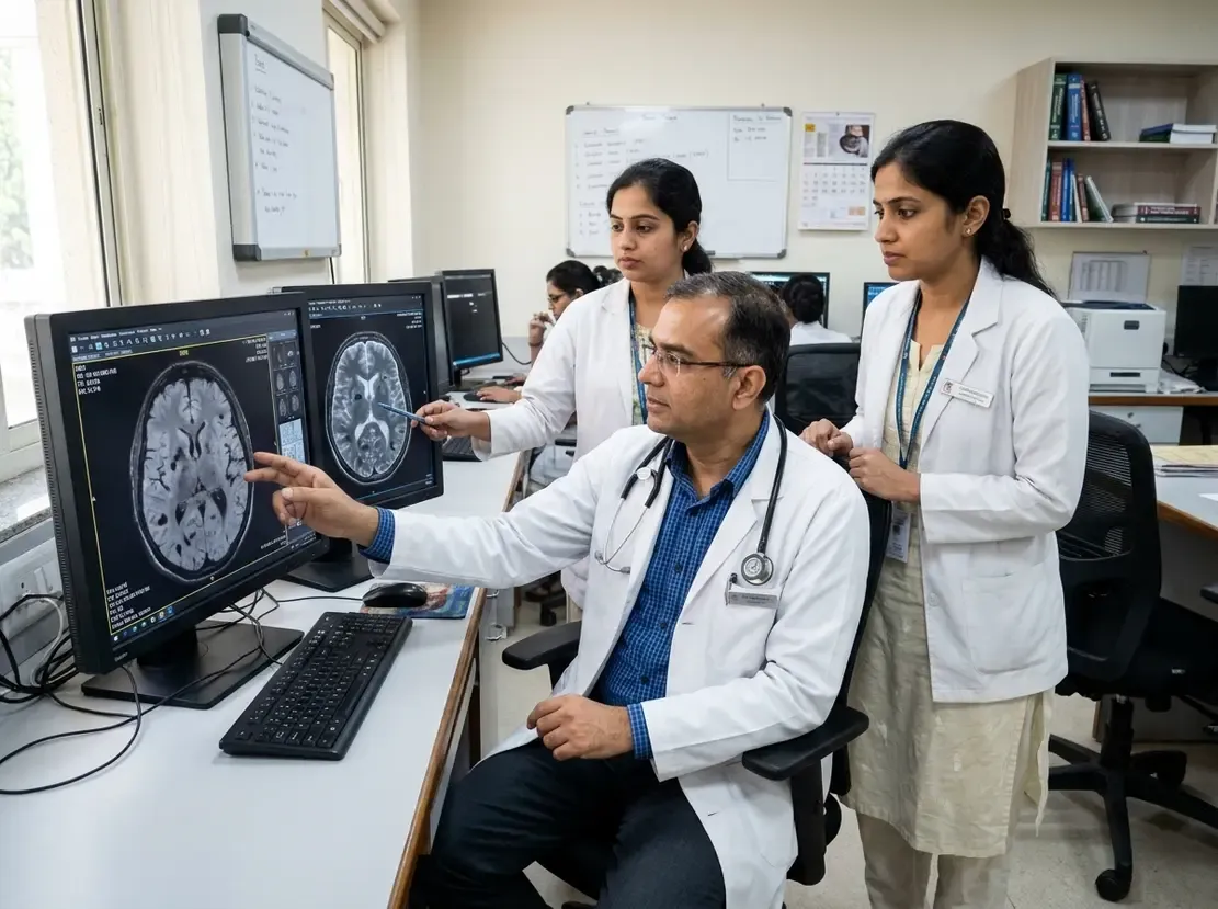
Modern magnetic resonance protocols rely heavily on susceptibility-weighted imaging to evaluate subtle intracranial abnormalities. This powerful technique detects paramagnetic and diamagnetic substances with exceptional precision. Consequently, clinicians obtain superior visualization of microscopic hemorrhage, deoxygenated venous blood, and pathological calcifications. Recently, European radiology societies released essential recommendations to guide standard acquisition and clinical interpretation.
Technical Essentials of Susceptibility-Weighted Imaging
To achieve diagnostic accuracy, teams must acquire high-resolution 3D gradient-echo data. Furthermore, clinicians should never evaluate magnitude images in isolation. Radiologists must systematically analyze four distinct image sets simultaneously. Specifically, these datasets include source magnitude images, filtered phase images, processed SWI maps, and minimum-intensity projection reconstructions.
In addition, interpreters must recognize blooming artefacts around dense structures. Vendor-specific phase conventions also demand careful attention during routine review. For instance, paramagnetic blood products and diamagnetic calcium produce opposite phase signals depending on the vendor software. Therefore, understanding local scanner display conventions prevents major misdiagnoses.
Core Brain Indications Supported by Clinical Evidence
Susceptibility-weighted imaging demonstrates its strongest clinical utility across specific brain disorders. First, specialists recommend this sequence for evaluating cerebral amyloid angiopathy. Moreover, the sequence provides critical baseline and longitudinal monitoring for amyloid-related imaging abnormalities with haemosiderin deposition during anti-amyloid therapy.
Additionally, neurotrauma protocols benefit greatly from this sensitive tool. SWI readily exposes subtle axonal shear injuries and petechial hemorrhages after traumatic brain injury. Furthermore, neuro-oncologists utilize these maps to assess intratumoral vascularity and internal calcification patterns.
In demyelinating disease, the sequence reliably detects the central vein sign and paramagnetic rim lesions. Consequently, these findings assist physicians in confirming multiple sclerosis diagnoses. Similarly, clinicians depend on the technique to map occult vascular malformations such as cavernomas and developmental venous anomalies.
Role of Susceptibility-Weighted Imaging Outside Neuroradiology
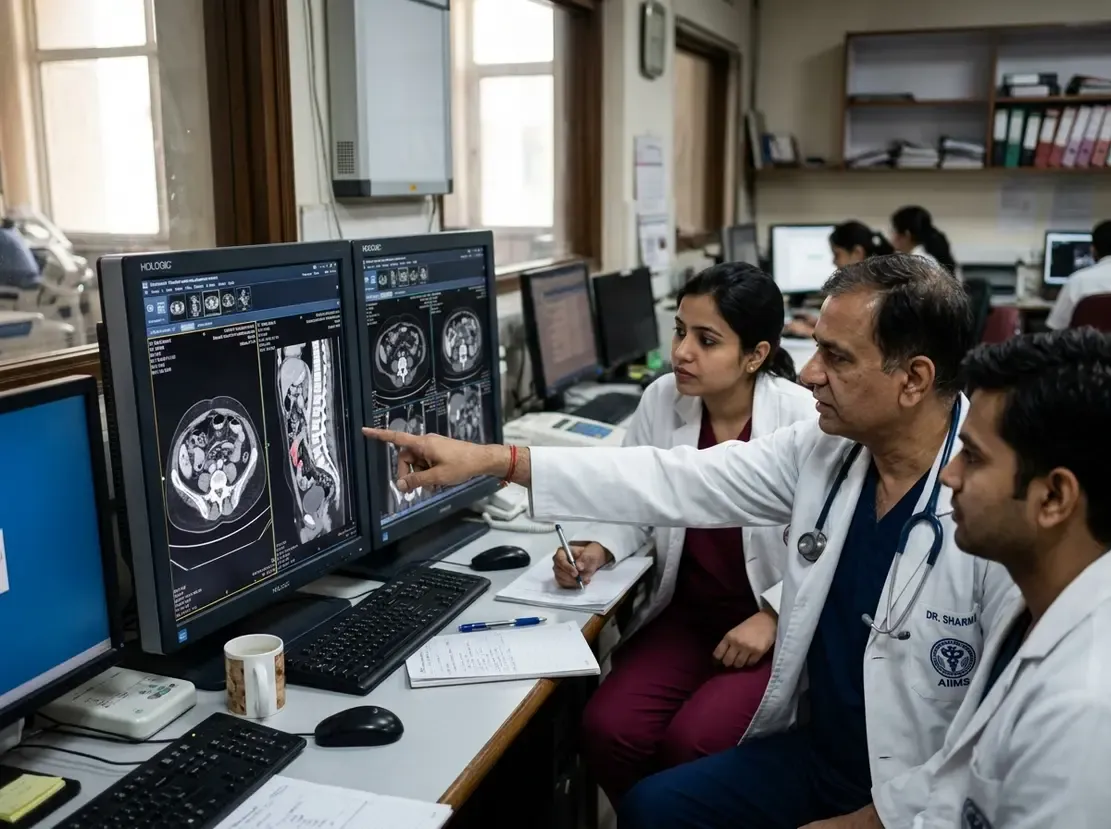
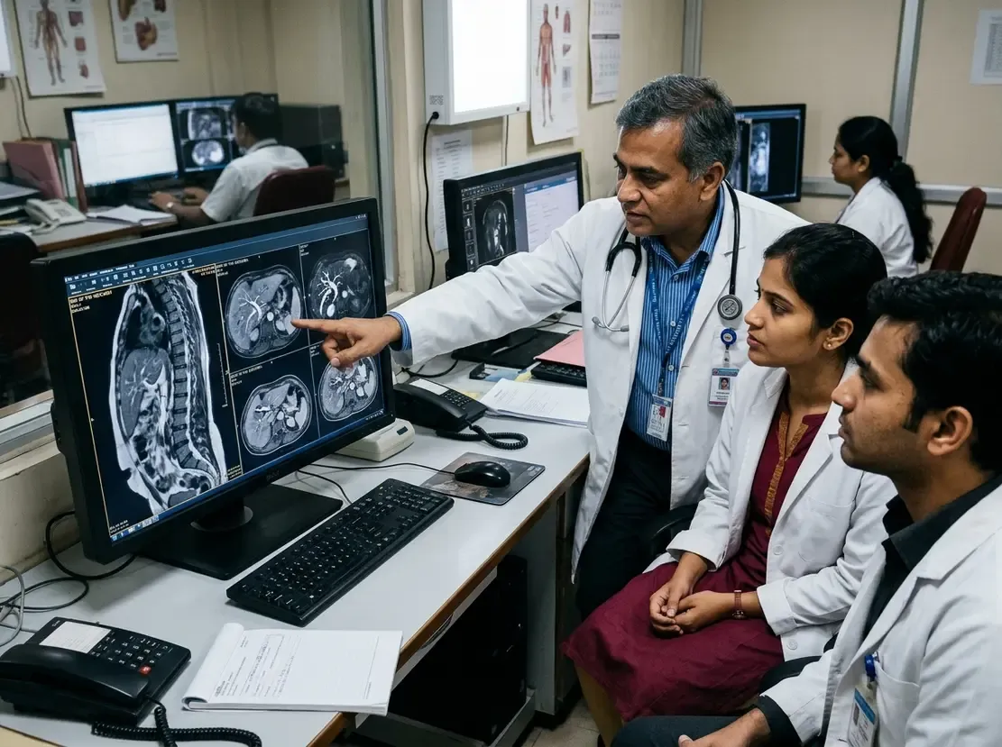
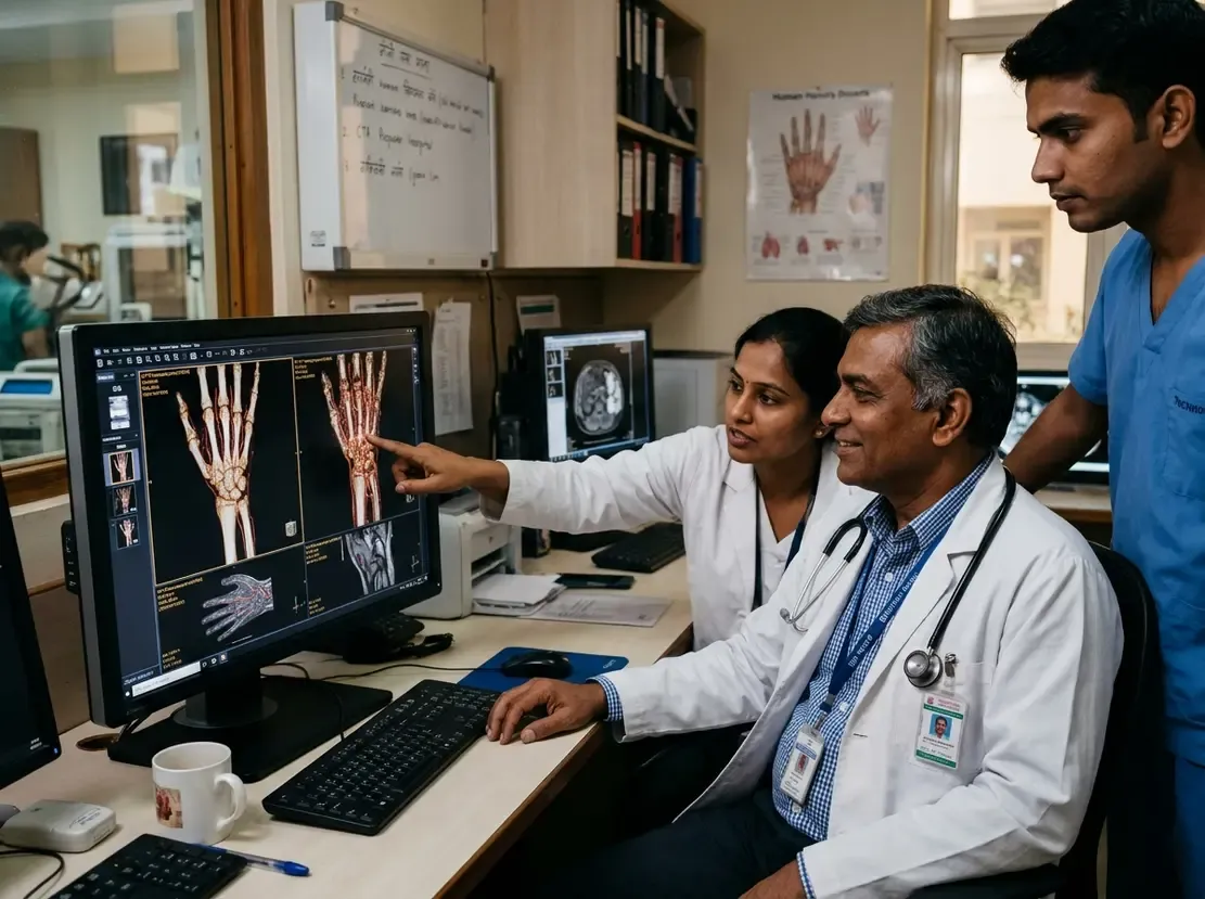
Currently, applications outside the central nervous system remain less standardized. However, specialists increasingly explore targeted uses in musculoskeletal, abdominal, breast, pelvic, and cardiac imaging. In these settings, teams should reserve the sequence for dedicated problem-solving rather than routine screening. Therefore, doctors should apply SWI when standard protocols cannot definitively characterize vascularity or hemorrhage.
Frequently Asked Questions
Q1: Why must clinicians inspect phase images alongside magnitude data in SWI?
Filtered phase images reliably differentiate paramagnetic substances like hemosiderin from diamagnetic mineral deposits like calcium. Furthermore, relying only on magnitude images can cause diagnostic confusion due to similar signal loss.
Q2: What role does SWI play during modern anti-amyloid antibody therapy?
Clinicians use SWI to identify amyloid-related imaging abnormalities with haemosiderin deposition, known as ARIA-H. Consequently, baseline and regular follow-up scans protect patients from severe hemorrhagic complications.
Q3: Should centers routinely add SWI to all non-neuro body MRI scans?
No, standard protocols outside neuroradiology do not require routine SWI. Instead, clinicians should reserve the sequence as a targeted problem-solving tool for ambiguous lesions.
References
- Parillo M et al. ESR Essentials: susceptibility-weighted imaging-practice recommendations by the European Society for Magnetic Resonance in Medicine and Biology. Eur Radiol. 2026 Oct 31. doi: 10.1007/s00330-026-12988-5. PMID: 42965692.
- Haller S, Haacke EM, Thurnher MM, Barkhof F. Susceptibility-weighted Imaging: Technical Essentials and Clinical Neurologic Applications. Radiology. 2021;299(1):19-35.
- Halefoglu AM, Yousem DM. Susceptibility weighted imaging: Clinical applications and future directions. World J Radiol. 2018;10(4):30-45.