Can 5.0 T MRI Transform Upper Abdominal Diagnostics?
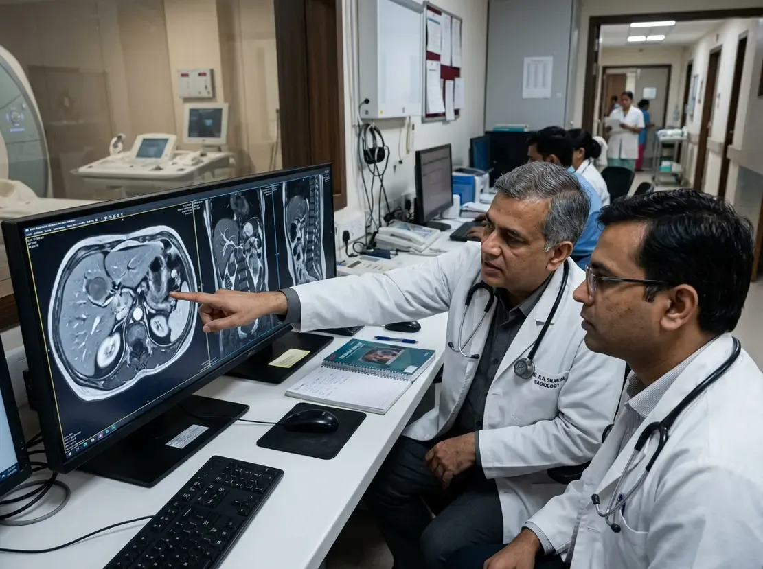
Advancements in High-Field Abdominal Imaging
Recent advances in ultra-high-field systems have established 5.0 T abdominal MRI as a promising diagnostic modality. Traditionally, clinicians rely on 1.5 T and 3.0 T systems for routine abdominal scans. However, higher magnetic fields offer substantial gains in signal and spatial resolution. Ultra-high-field imaging historically presented technical challenges, such as dielectric shading and elevated energy absorption. Consequently, body imaging at fields above 3.0 T required careful engineering solutions. A landmark prospective study now provides robust evidence confirming the clinical feasibility of 5.0 T imaging.
Comparing Field Strengths in Upper Abdominal Scans
Researchers evaluated both a prospective patient cohort and a healthy volunteer cohort. Specifically, the patient group included 348 consecutive individuals undergoing upper abdominal examinations. In addition, 31 healthy volunteers underwent scans across all three magnetic field strengths. Two independent radiologists assessed subjective parameters, including anatomical detail, artifact burden, and diagnostic confidence. Furthermore, the investigators calculated objective quantitative metrics, such as signal-to-noise ratio and contrast-to-noise ratio.
Clinical Performance of 5.0 T Abdominal MRI
The objective quantitative analysis demonstrated significant advantages for the ultra-high-field system. Specifically, measurements revealed a clear gradient where 5.0 T surpassed both 3.0 T and 1.5 T. Liver signal-to-noise ratio, spleen SNR, and muscle SNR increased markedly at 5.0 T. Moreover, the liver-to-spleen contrast-to-noise ratio showed superior values on the 5.0 T scanner. Subjective ratings also confirmed excellent delineation of parenchymal structures and vascular anatomy. Therefore, radiologists reported exceptionally high diagnostic confidence without prohibitive artifacts.
Relevance for Abdominal Radiology
These findings carry direct relevance for advanced cross-sectional imaging in modern radiology centers. Early detection of subtle hepatic lesions and microvascular invasion requires optimal spatial resolution and contrast. In addition, higher signal allows clinicians to optimize scan duration while maintaining image clarity. While 1.5 T and 3.0 T remain workhorses across many hospitals, 5.0 T technology bridges ultra-high-field physics with daily clinical needs. Thus, adopting these protocols may significantly improve patient workup in complex hepatobiliary diseases, supported by specialized training in gastro intestinal radiology for diagnostic accuracy.
Frequently Asked Questions
Q1: What are the primary advantages of 5.0 T abdominal MRI over lower field strengths?
The 5.0 T platform provides substantially higher signal-to-noise and contrast-to-noise ratios compared to 1.5 T and 3.0 T systems. Consequently, radiologists achieve sharper anatomical delineation of liver structures and vascular details, which improves overall diagnostic confidence.
Q2: Did the 5.0 T scans suffer from severe dielectric or motion artifacts?
The prospective study demonstrated that modern sequence optimization effectively manages artifacts. In fact, radiologists rated the overall image quality at 5.0 T consistently high, confirming that artifact control is clinically acceptable for upper abdominal evaluations.
Q3: How do the quantitative SNR and CNR measurements compare across the scanners?
Objective data showed a statistically significant progressive gradient: 5.0 T exceeded 3.0 T, and 3.0 T exceeded 1.5 T. This improvement was evident in liver SNR, splenic SNR, and liver-to-spleen CNR across both cohorts, aligning with advanced curricula found in radiology speciality courses.
References
- Zhu L et al. 5.0 T versus 1.5 T and 3.0 T upper abdominal MRI: a prospective two‑cohort study of image quality and diagnostic confidence. Eur Radiol. 2026 Sep 28. doi: 10.1007/s00330-026-12874-0. PMID: 42803937.
- Zhao H, Xu Q, Gao R, et al. Clinical Feasibility of 5.0 T MRI/MRCP in Characterizing Pancreatic Cystic Lesions: Comparison with 3.0 T and MDCT. Diagnostics (Basel). 2024;14(21):2440.
- Yin L, Li Z, Shang M, et al. Magnetic resonance cholangiopancreatography at 5.0 T: quantitative and qualitative comparison with 3.0 T. BMC Med Imaging. 2024;24(1):331.