AI Brain Age Estimation Shows Promise in Dementia Care
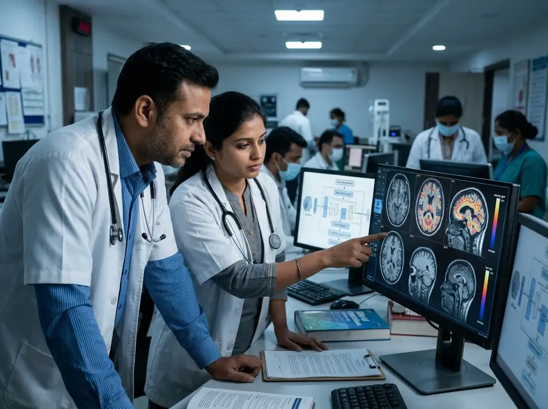
Clinicians are increasingly exploring artificial intelligence to evaluate structural neuroimaging scans. Specifically, brain age estimation provides an objective biomarker of biological brain health. Researchers recently evaluated a practical deep learning pipeline using standard T1-weighted magnetic resonance imaging. Consequently, this tool accurately predicts chronological age and reveals critical neurodegenerative risks.
Framework and Accuracy of Brain Age Estimation
First, the research team trained the model on 4,346 cognitively normal adults. Notably, the engineers relied exclusively on freely accessible Python tools for image preprocessing. This computational approach avoids cumbersome software suites that delay clinical workflows. Furthermore, the model demonstrated an impressive correlation of 0.95 with chronological age. Additionally, the internal test set yielded a mean absolute error of only 3.76 years. Meanwhile, external test cohorts achieved mean absolute errors between 4.82 and 5.15 years. Therefore, the framework maintains dependable accuracy across independent hospital datasets.
Clinical Significance of the Brain-Age Gap
Clinicians define the brain-age gap as predicted biological age minus actual chronological age. Importantly, an elevated residualized gap indicates accelerated brain aging. The researchers evaluated this biological marker across 954 patients in the BICWALZS cohort. As expected, patients with neurodegenerative dementias exhibited significantly higher brain-age gaps. Furthermore, positive amyloid PET scans correlated strongly with widened age discrepancies. In addition, elevated brain-age gaps aligned with severe white matter hyperintensities and prior strokes. Consequently, longitudinal analysis confirmed that higher gaps independently predict clinical worsening through dedicated stroke medicine evaluations.
Relevance for Neurological Practice in India
Dementia cases are escalating rapidly across aging populations in India. However, advanced molecular imaging like amyloid PET remains expensive and scarce in many centers. In contrast, T1-weighted MRI scanners operate widely in Indian diagnostic centers. Because open-source Python tools require minimal computational power, local clinics can adopt this pipeline. Thus, physicians can stratify dementia risk without imposing excessive financial burdens on families. Ultimately, rapid automated triage helps neurologists prioritize patients who need intensive early intervention.
Frequently Asked Questions
Q1: What does brain age estimation measure on an MRI?
It estimates biological brain age by analyzing structural patterns and tissue atrophy on T1-weighted MRI scans. Furthermore, it compares this value against normal aging trajectories.
Q2: Why is the brain-age gap clinically useful?
A positive brain-age gap indicates accelerated neurodegeneration. Moreover, studies link this gap directly to amyloid pathology, vascular injury, and faster cognitive decline.
Q3: Can community diagnostic clinics implement this AI tool?
Yes, because the framework relies on freely available Python packages for preprocessing. Therefore, centers can run rapid evaluations on standard hardware without proprietary software.
References
- Kim HW et al. Evaluation of a Rapid and Practical Deep Learning Framework for T1-weighted MRI-based Brain Age Estimation. Radiol Artif Intell. 2026 Nov 11. doi: 10.1148/ryai.260310. PMID: 43012635.
- Kaufmann T, et al. Common brain disorders are associated with heritable patterns of apparent aging of the brain. Nat Neurosci. 2019;22(10):1617-1623.
- Jones DT, et al. Deep learning-based brain age prediction in normal aging and dementia. Nat Aging. 2022;2(5):412-424.