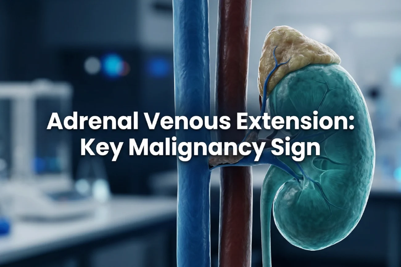
Surgeons often encounter Adrenal Venous Extension during the management of malignant lesions. This complication affects both primary adrenocortical carcinomas and metastatic tumors. Consequently, understanding the imaging markers that predict such involvement remains vital for improving patient outcomes. In India, where adrenal incidentalomas are increasingly detected, accurate staging via cross-sectional imaging is essential for therapeutic success.
Prevalence of Adrenal Venous Extension
Recent data indicates that venous involvement occurs frequently in malignant adrenal neoplasia. For instance, approximately 21.4% of patients with adrenocortical carcinoma (ACC) present with this finding. Meanwhile, 14.6% of patients with adrenal metastases also exhibit venous extension. Although ACC shows a slightly higher tendency for vascular invasion, the difference between primary and secondary tumors is not statistically significant. Therefore, radiologists must remain vigilant when assessing any malignant adrenal mass regardless of its origin. Furthermore, lung, colorectal, and breast cancers represent the most common primary sources for these metastatic lesions.
Role of the Edge Sign in Imaging
The “edge sign” represents a novel morphological indicator that may precede actual Adrenal Venous Extension. In metastatic cases, this sign appears in nearly 26.8% of lesions compared to 17.8% in primary carcinomas. Additionally, multivariate analysis suggests that tumor size and the edge sign independently predict extension into the renal or adrenal veins. Thus, identifying this feature early allows for more precise diagnostic staging. Because the edge sign provides a visual warning of impending invasion, it serves as a critical tool for the radiology department. Moreover, interobserver agreement for this sign remains substantial among experienced readers.
Clinical Relevance and Surgical Planning
These findings could fundamentally change how doctors manage adrenal cancer. Because the edge sign identifies early extension, it helps prioritize patients for surgical intervention. In addition, recognizing these patterns aids in distinguishing between different types of adrenal neoplasia during the workup. Specifically, clinicians can use these markers to expedite surgery and prevent further vascular complications. However, practitioners should wait for prospective multicenter studies to fully confirm the clinical impact of these imaging signs. Consequently, incorporating these observations into routine reports could enhance the precision of oncological care.
Frequently Asked Questions
Q1: What is the “edge sign” in adrenal imaging?
The edge sign refers to specific morphological changes in tumor shape that often indicate early venous involvement. It effectively serves as a precursor to actual vascular extension in malignant adrenal lesions.
Q2: How common is venous extension in metastatic adrenal cancer?
Research shows that venous extension occurs in about 14.6% of metastatic adrenal lesions. These cases predominantly involve primary cancers originating from the lungs or colorectal system.
Q3: Does tumor size affect the risk of venous extension?
Yes, mean tumor size is a strong independent predictor of extension into the adrenal and renal veins. Larger tumors generally carry a higher risk of vascular invasion.
References
- Melges LPDM et al. Prevalence of venous extension in malignant adrenal neoplasia: beyond primary tumors and identification of a novel imaging sign. Eur Radiol. 2026 Feb 28. doi: 10.1007/s00330-026-12382-1. PMID: 41762262.
- Boland GW. Adrenal Imaging: From Basic Interpretation to Advanced Evaluation. RadioGraphics. 2004;24 Spec No:S87-105.
- Hussain S, et al. Adrenocortical Carcinoma: The Range of Appearances on CT and MRI. AJR Am J Roentgenol. 2018;210(4):W141-W151.