How ASCVD Imaging Biomarkers Revolutionize Heart Health
Atherosclerotic cardiovascular disease remains the leading cause of global mortality. Consequently, early detection during the prolonged subclinical phase offers vital opportunities for timely intervention. Traditional risk calculation tools often misclassify risk in borderline or intermediate patients. Therefore, clinicians increasingly rely on ASCVD imaging biomarkers to guide personalized preventive therapies. These imaging biomarkers identify subclinical atherosclerosis directly before clinical events occur.
Role of ASCVD Imaging Biomarkers in Risk Assessment
Coronary artery calcium scoring provides an established tool for cardiovascular risk reclassification. Specifically, noncontrast electrocardiographically gated computed tomography measures coronary artery calcium accurately. In addition, zero calcium scores identify individuals who can safely defer statin treatment. However, elevated scores highlight individuals who need intensive lipid-lowering medical therapy. Thus, direct visualization of calcified plaque significantly refines risk prediction across asymptomatic populations.
Advanced Coronary Plaque Characterization
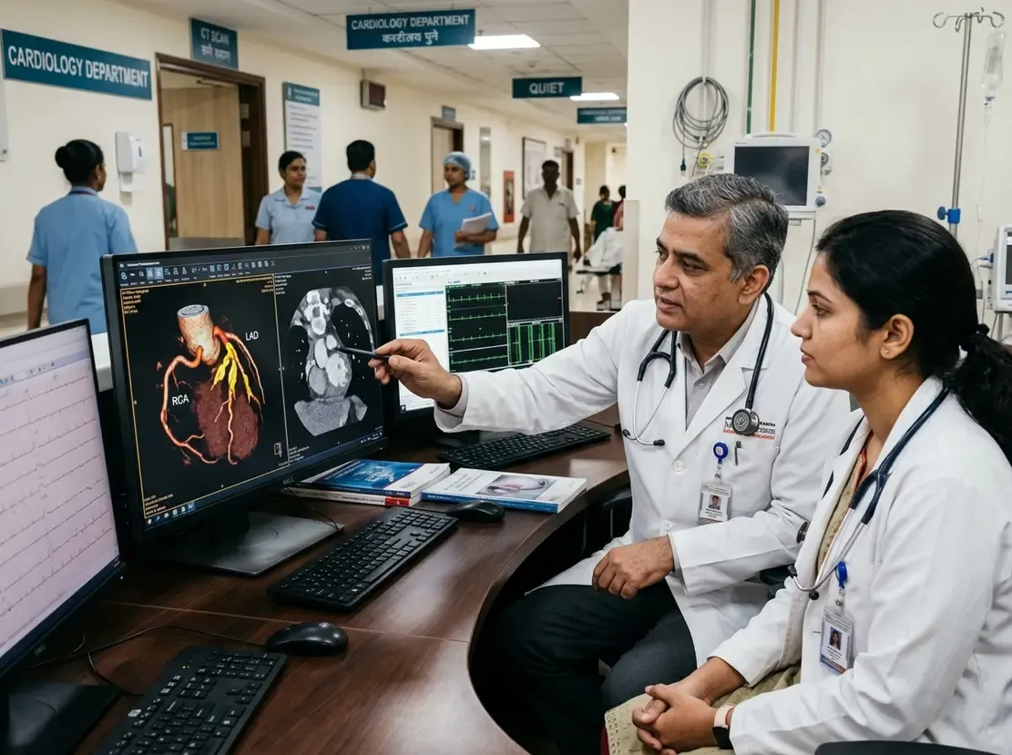
Coronary CT angiography offers detailed anatomical evaluation beyond standard calcium scoring. For instance, it evaluates overall plaque burden and plaque composition with great precision. Furthermore, advanced angiography detects high-risk plaque features like low-attenuation plaque and positive remodeling. Moreover, quantitative plaque analysis and pericoronary adipose tissue imaging reveal vascular inflammatory activity. Consequently, these comprehensive parameters help clinicians identify unstable disease before acute coronary events arise.
Extracoronary and Opportunistic Imaging Biomarkers
Cardiovascular screening extends beyond the coronary arteries. For example, carotid and aortic ultrasound assess systemic vascular stiffness and atherosclerosis. In addition, epicardial adipose tissue measurement provides valuable metabolic insights. Routine chest and abdominal imaging examinations also yield valuable opportunistic biomarkers without extra radiation exposure. Specifically, radiologists can evaluate incidental vascular calcifications, breast arterial calcifications, and hepatic steatosis. Similarly, CT scans reveal visceral adiposity, sarcopenia, and osteoporosis during regular patient assessments. Ultimately, integrating artificial intelligence and standardized reporting into clinical workflows will expand personalized cardiovascular prevention.
Frequently Asked Questions
Q1: Why are imaging biomarkers important in cardiovascular screening?
Imaging biomarkers directly quantify subclinical arterial plaque. Therefore, they refine individual risk stratification far better than traditional risk calculators alone.
Q2: What is the most established coronary imaging biomarker for screening?
Coronary artery calcium scoring remains the most guideline-endorsed biomarker. Specifically, it provides robust prognostic information without requiring contrast dye.
Q3: How do opportunistic imaging biomarkers benefit patients?
Opportunistic biomarkers identify incidental vascular calcifications and visceral adiposity on routine scans. Consequently, patients gain cardiovascular insights without receiving additional radiation.
References
- Sorin V et al. State-of-the-Art Imaging Biomarkers in Screening of Atherosclerotic Cardiovascular Disease. Radiographics. 2026 Oct undefined. doi: 10.1148/rg.260068. PMID: 42784472.
- Darimireddi A, Panigrahi N, Golive A, Kodem S, Gudivada J. Imaging and Biomarkers for Subclinical Atherosclerosis in Women. Indian J Cardiovasc Dis Women. 2025;10:294-303. doi: 10.25259/IJCDW_58_2025.
- den Dekker MAM. Imaging biomarkers for detection of coronary artery disease. University of Groningen; 2014.