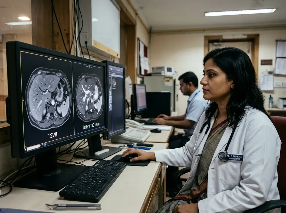
Predicting Post-TIPS Encephalopathy with 3D Body Scans
Transjugular intrahepatic portosystemic shunt decompresses the portal system in cirrhotic patients. However, post-TIPS hepatic encephalopathy remains a devastating clinical complication that limits procedural utility. Traditional clinical risk scores offer modest predictive accuracy. Therefore, clinicians urgently require objective biomarkers to identify high-risk individuals prior to shunt placement.
Limitations of 2D Body Composition Metrics
Sarcopenia and skeletal muscle depletion strongly correlate with neurocognitive decline in cirrhosis. Historically, radiologists evaluated body composition by manually measuring muscle area on a single L3 axial slice. Nevertheless, manual two-dimensional measurements display severe interobserver variability. In fact, recent data reveal significant measurement variation across 96.2% of two-dimensional features. Consequently, standard single-slice cross-sectional imaging provides inadequate predictive value for shunt candidates.
Predicting Post-TIPS Hepatic Encephalopathy with 3D AI
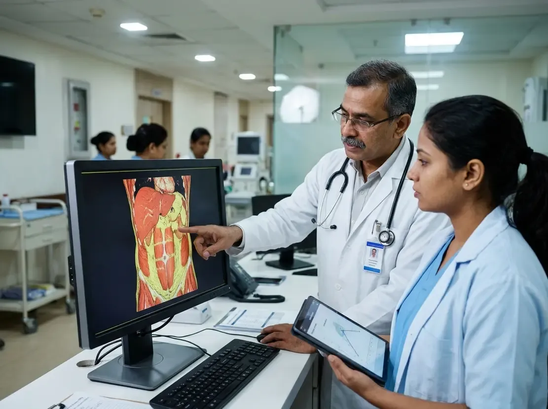
To overcome these limitations, investigators engineered a fully automated three-dimensional evaluation framework. Specifically, the researchers developed volumetric segmentation algorithms that evaluate whole-abdomen muscle and adipose tissue. Furthermore, this platform extracts comprehensive morphologic and radiomic features from routine abdominal computed tomography scans. The study analyzed 593 cirrhotic patients across seven clinical centers to assess post-TIPS hepatic encephalopathy risk. Importantly, the multi-institutional cohort included both internal training and two independent external validation cohorts.
Superior Performance of the Integrated Model
The novel three-dimensional model substantially outperformed conventional two-dimensional manual metrics. In addition, researchers combined the volumetric data with key clinical variables to construct an integrated model. This integrated system achieved impressive area under the curve values of 0.78, 0.73, and 0.77 across cohorts. By comparison, traditional prognostic tools like Child-Pugh, MELD, and ALBI achieved area under the curve values below 0.65. Thus, three-dimensional radiomic profiling provides markedly superior discrimination for post-procedural neurocognitive impairment.
Clinical Implications for Hepatology Practice
Preoperative risk stratification plays an essential role in optimizing outcomes after portal decompression. For example, interventional teams can tailor shunt diameter or adopt prophylactic measures for high-risk candidates. Moreover, automated software pipelines eliminate interobserver variability and integrate seamlessly into diagnostic workflows. Ultimately, volumetric body composition assessment promises to refine patient selection and safeguard post-shunt clinical recovery.
Frequently Asked Questions
Q1: Why do standard 2D CT measurements fail to reliably predict overt hepatic encephalopathy?
Single-slice two-dimensional measurements show marked interobserver variability and fail to capture full volumetric muscle and fat architecture. Consequently, they miss subtle radiomic changes that indicate systemic metabolic vulnerability.
Q2: How does the automated 3D model improve upon conventional clinical scores like MELD?
Traditional scores like MELD and Child-Pugh evaluate end-organ liver and renal dysfunction rather than metabolic reserve. In contrast, the integrated 3D model quantifies volumetric body composition and radiomic signatures, achieving superior predictive discrimination.
Q3: Can clinicians readily integrate automated 3D body composition analysis into routine care?
Yes, because the automated algorithm processes standard pre-procedural abdominal computed tomography scans without requiring extra imaging visits. Therefore, teams can seamlessly calculate individualized risk metrics during routine pre-operative workups.
References
- Zhao Y et al. Three-dimensional automated muscle and adipose analysis to predict hepatic encephalopathy after transjugular intrahepatic portosystemic shunt. Eur Radiol. 2026 Nov 15. doi: 10.1007/s00330-026-13063-9. PMID: 43034938.
- Mukund A, Bhardwaj V, Jindal A, Patidar Y, Sarin SK. Transjugular Intrahepatic Portosystemic Shunt Related Hepatic Encephalopathy in Cirrhotics With Refractory Ascites: Incidence and Correlation With TIPS Stent Diameter and Pre-TIPS Sarcopenia. J Clin Exp Hepatol. 2021;11(6):663-671.
- Chen X, Wang T, Ji Z, et al. 3D automatic liver and spleen assessment in predicting overt hepatic encephalopathy before TIPS: a multi-center study. Hepatol Int. 2023;17(6):1545-1556.