Can AI Accurately Assess Mammographic Breast Density?
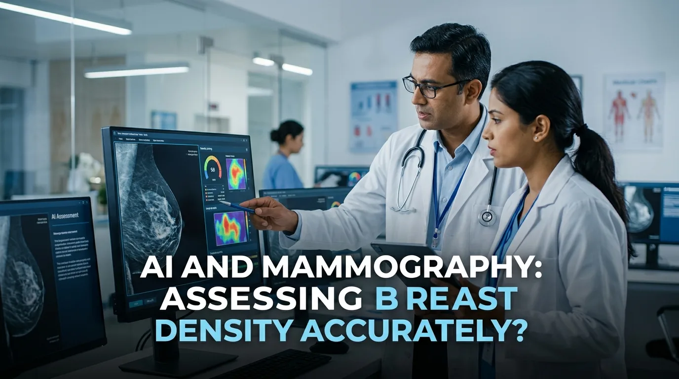
Dense breast tissue lowers mammography sensitivity and significantly increases cancer risk. Consequently, implementing an automated breast density assessment using artificial intelligence (AI) has emerged as a promising solution to standardize risk-stratified screening. However, recent real-world evidence indicates that vendor-specific variations and anatomical discrepancies present important clinical hurdles.
Understanding Automated Breast Density Assessment in Screening
A recent large-scale study evaluated 200,000 screening mammograms from BreastScreen Norway to analyze AI-driven volumetric breast density (VBD) assessment and cancer risk scores. The researchers categorized continuous VBD into four distinct density tiers (VBD 1 through 4). Furthermore, they analyzed malignancy detection accuracy using receiver operating characteristic curves across different imaging vendors.
Key Findings and Diagnostic Performance
Overall, the commercial AI algorithm demonstrated robust cancer detection performance across all density strata. However, diagnostic accuracy decreased steadily as breast density increased. Specifically, the area under the curve (AUC) was 0.955 for fatty breasts (VBD 1), but dropped to 0.857 for extremely dense breasts (VBD 4).
In addition, the distribution of high-density classifications varied markedly between different mammography equipment manufacturers. For instance, Vendor B classified twice as many exams into the highest density category (6.7%) compared to Vendor A (3.3%). Moreover, 18.5% of examinations exhibited different density categories between the right and left breasts of the same patient.
Clinical Implications for Practice
These findings highlight both opportunities and caveats for clinical radiology workflows. Therefore, clinicians must exercise caution before relying solely on automated density metrics to recommend supplemental ultrasound or MRI. While AI offers rapid volumetric quantification, healthcare systems must account for vendor-dependent calibration and inter-breast variations before implementing standardized screening protocols.
Frequently Asked Questions
Q1: Why does mammographic sensitivity decrease in dense breasts?
Dense fibroglandular tissue appears white on mammograms, which creates a masking effect that can obscure small tumors and calcifications.
Q2: How does AI improve breast density evaluation?
AI tools provide automated volumetric quantification, which reduces inter-radiologist subjectivity and helps standardize risk assessment.
Q3: What challenges remain before widespread clinical adoption?
Differences across equipment vendors and bilateral breast variations require careful calibration before deploying AI density models into routine screening pathways.
References
- Larsen M et al. Mammographic density assessment by an artificial intelligence model for breast cancer detection in BreastScreen Norway. Eur Radiol. 2026 Aug 13. doi: 10.1007/s00330-026-12786-z. PMID: 42593501.
- Lång K et al. Artificial intelligence-supported screen reading versus standard double reading in the Mammography Screening with Artificial Intelligence trial (MASAI): a clinical safety analysis of a randomised, controlled, non-inferiority, single-blinded, screening accuracy study. Lancet Oncol. 2023;24(8):868-876.
- Gastounioti A et al. Evaluation of an artificial intelligence model for longitudinal consistency in mammographic breast density assessment. BJR Artif Intell. 2025;2(1):ubaf004.