Can Routine Non-Head CT Scans Guide Cochlear Implants?
Significance of Automatic Cochlear Morphometry in Surgical Planning
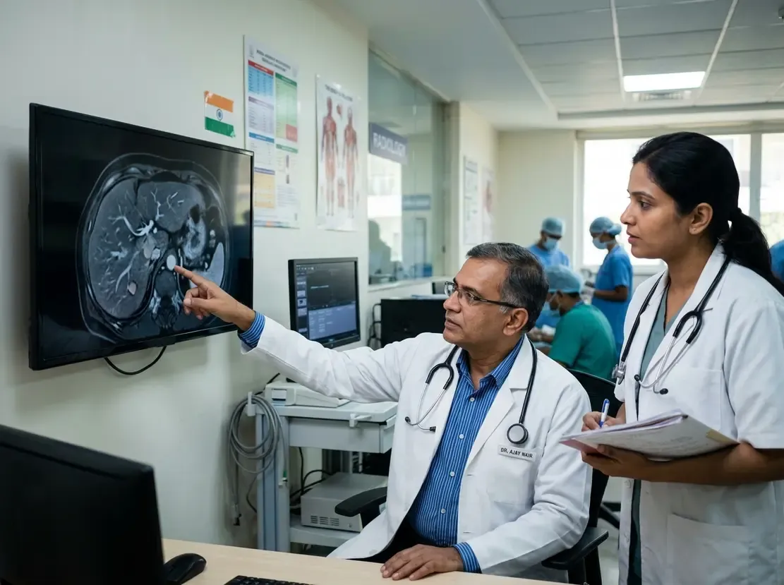
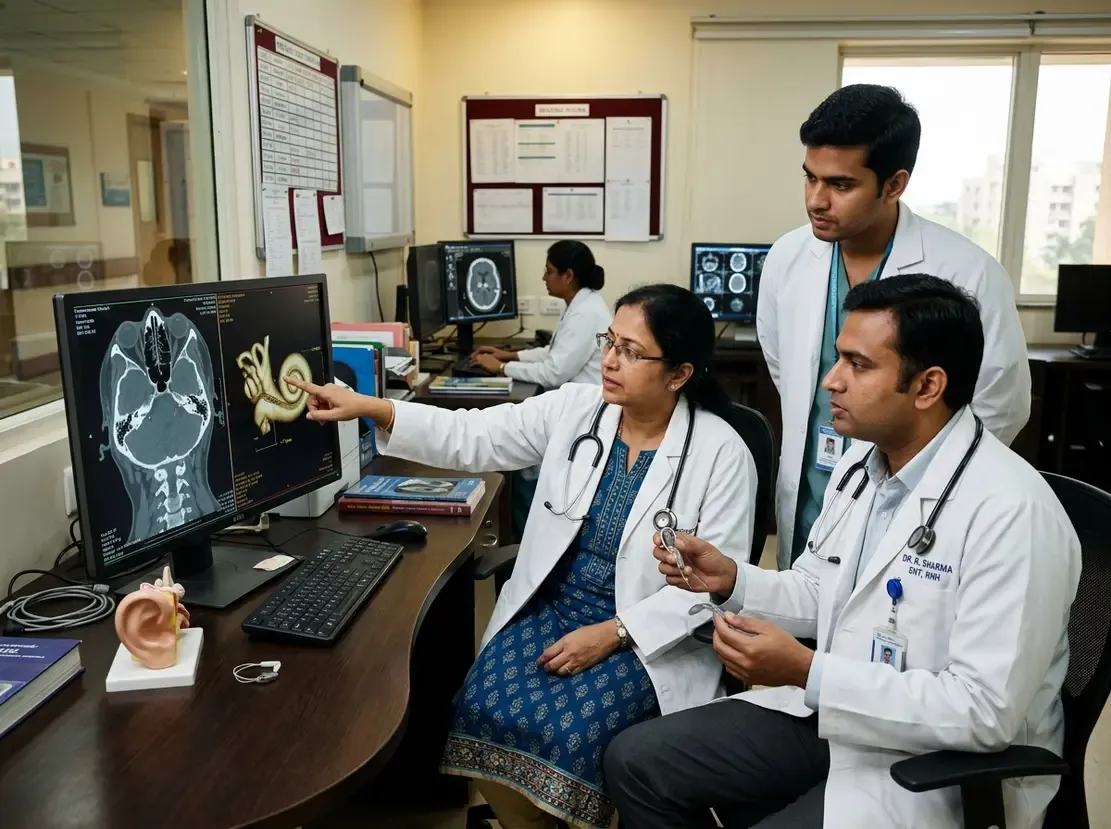
Precise electrode selection directly impacts hearing outcomes in cochlear implant candidates. Therefore, otologists frequently rely on automatic cochlear morphometry to estimate cochlear duct length accurately. However, standard temporal bone and head CT protocols often deliver high radiation doses to vulnerable ocular tissues. A recent multi-cohort study examined whether alternative head and neck CT scans provide reliable morphometric data. Consequently, this investigation evaluated if existing scans can replace dedicated high-dose head examinations. Clinicians often encounter patients who already have cervical spine, sinus, or carotid angiograms.
Comparing Head CT with Alternative CT Protocols
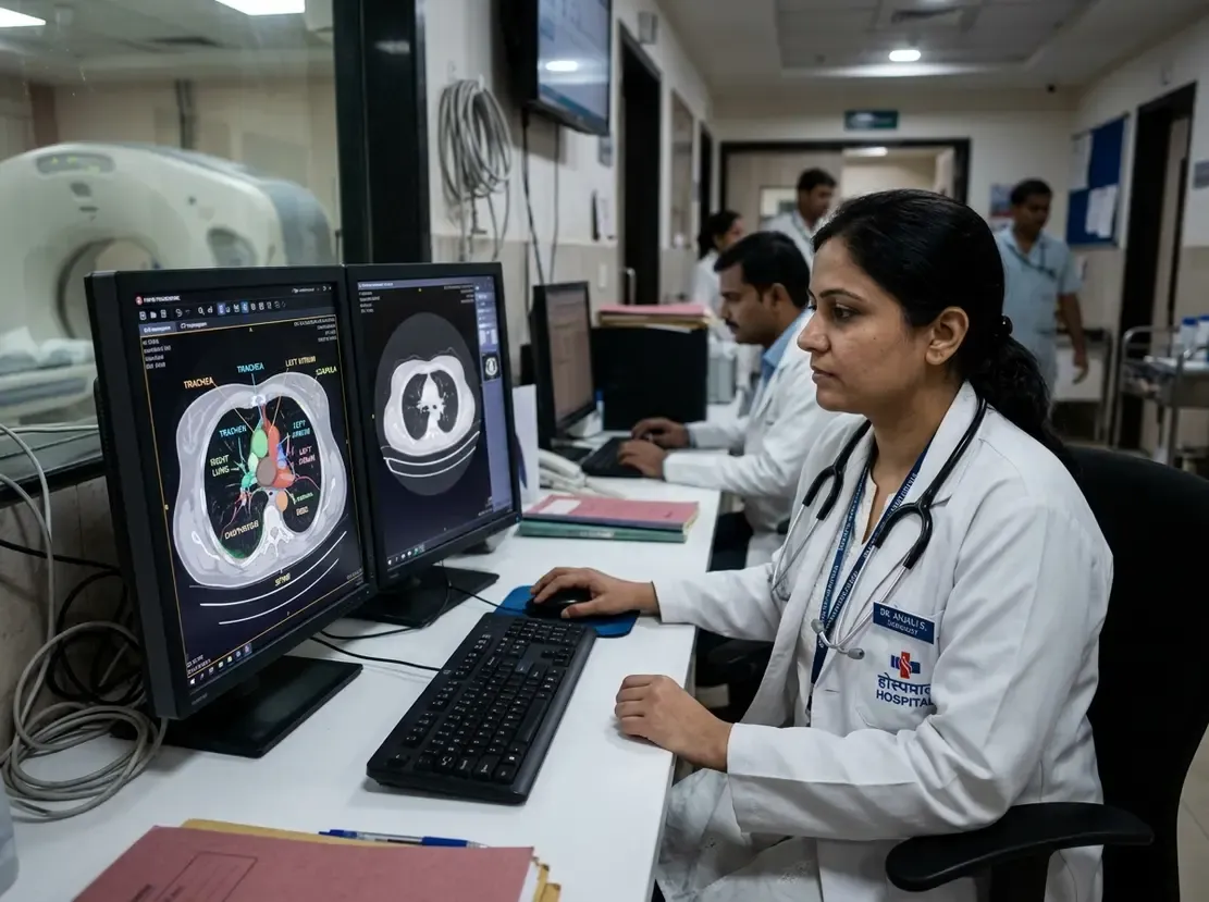
Researchers compared 459 intraindividual pairs across different protocol types. Specifically, the team assessed cervical spine CT, carotid CT angiography, and paranasal sinus CT against standard head CT. Standard head CT delivered a median radiation dose index of 53.2 mGy. In contrast, cervical spine CT exposed patients to only 16.3 mGy. Furthermore, carotid CT angiography and sinus CT delivered just 8.4 mGy and 5.0 mGy, respectively. Despite marked differences in radiation and contrast levels, cochlear duct length measurements demonstrated high intraindividual consistency. Thus, alternative neck and sinus scans successfully preserved anatomical precision for the organ of Corti.
Clinical Implications for Practice in India
In Indian clinical settings, surgical candidates often present with pre-existing diagnostic scans from varied indications. For example, trauma victims or elderly individuals frequently undergo carotid or cervical spine imaging during acute triage. Consequently, re-using these incidental scans prevents duplicate examinations and lowers overall treatment costs. Moreover, pediatric candidates under national cochlear implant initiatives benefit tremendously from lower radiation exposure. Otologists can confidently leverage automated software on existing scans to choose tailored electrode arrays. As a result, surgical teams can streamline preoperative workflows while strictly safeguarding patient safety.
Frequently Asked Questions
Q1: What is automatic cochlear morphometry in cochlear implant planning?
It is a software-driven process that calculates cochlear duct dimensions. Specifically, this helps surgeons select the optimal electrode length for each patient.
Q2: Can cervical spine or carotid CT replace head CT for cochlear measurements?
Yes, research shows these scans provide comparable cochlear duct length estimates. Furthermore, they deliver significantly lower radiation exposure than head CT.
Q3: How does this approach benefit clinical practice in India?
It lowers medical expenses by avoiding repeat scans. In addition, it protects pediatric patients from unnecessary cumulative radiation.
References
- Huflage H et al. Automatic cochlear morphometry: intraindividual comparison of head CT vs carotid CT angiography, cervical spine, and paranasal sinus CT. Eur Radiol. 2026 Sep 15. doi: 10.1007/s00330-026-12871-3. PMID: 42742655.
- Huflage H, Wech T, Rak K, et al. Influence of CT Radiation Dose and Field-of-View on Automatic Morphometry for Cochlear Implant Planning. Otol Neurotol. 2025;46(7):e250-e256.
- Alshalan A, et al. Radiation-free automatic planning for cochlear implantation: Comparing cochlear duct lengths between CT and MRI. Braz J Otorhinolaryngol. 2025;91(4):101582.