AI Foundation Models for Faster Breast MRI Triaging
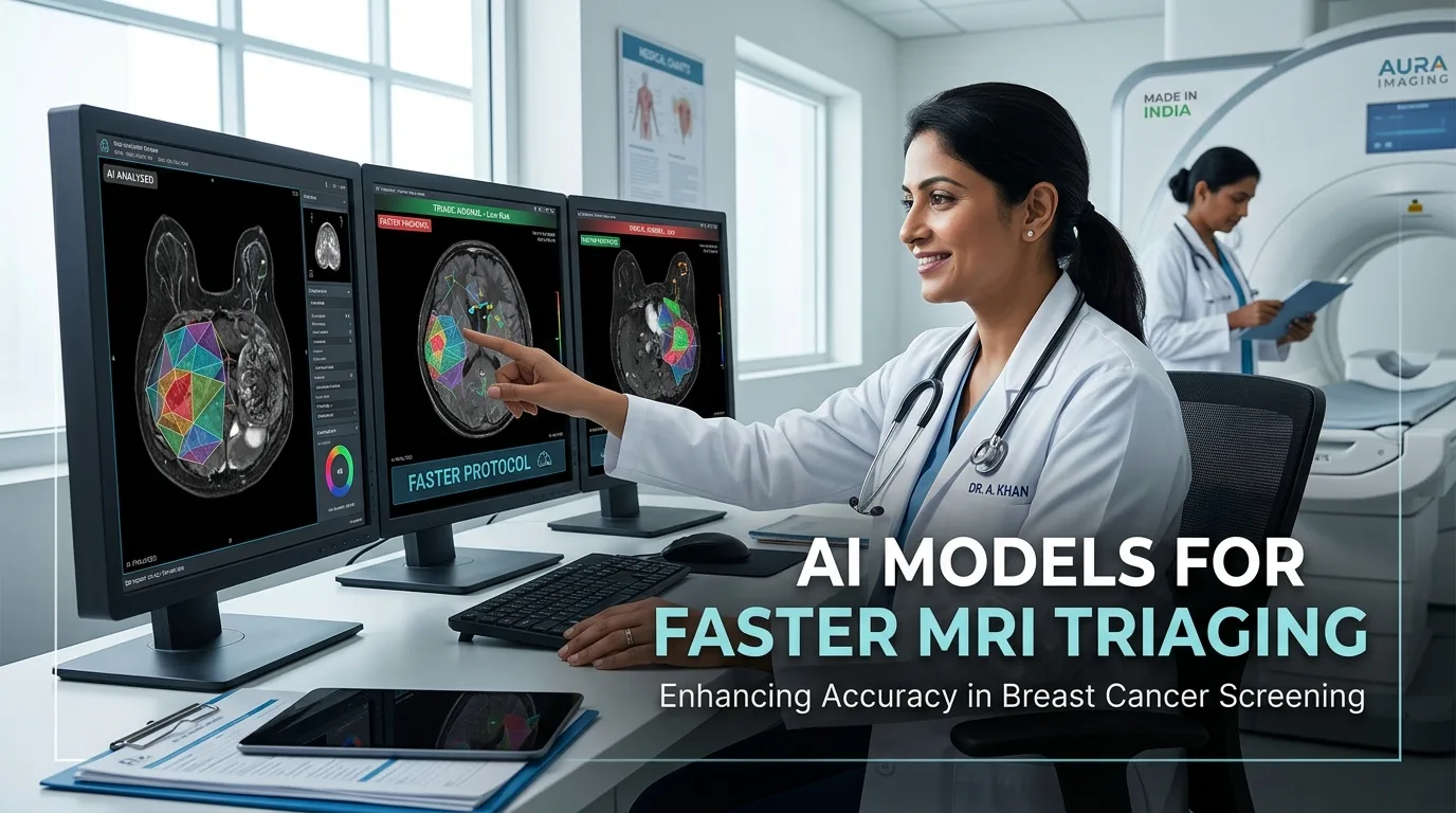
Abbreviated magnetic resonance imaging provides exceptional diagnostic sensitivity for detecting breast cancer. However, high interpretation workload limits its widespread clinical implementation. Recent developments in artificial intelligence now enable effective breast MRI triaging to rule out normal examinations rapidly. Consequently, clinicians can prioritize suspicious cases requiring urgent intervention.
Evaluating AI in Breast MRI Triaging
Researchers evaluated a medical slice transformer based on the DINOv2 vision foundation model. Specifically, the study analyzed 1,847 breast MRI examinations from an in-house cohort and 924 external scans. The team compared four distinct abbreviated imaging protocols. These included contrast-enhanced T1-weighted subtraction, diffusion-weighted imaging, and combinations with T2-weighted imaging. Moreover, the investigators assessed model accuracy at high sensitivity thresholds to mirror real clinical screening needs. Therefore, this evaluation provides robust evidence regarding automated triage capabilities.
Key Diagnostic Performance and Protocol Comparison
The model demonstrated strong discriminative performance across both contrast and non-contrast protocols. For instance, the combination of T1 subtraction and T2-weighted imaging achieved an AUC of 0.77. Similarly, non-contrast diffusion-weighted imaging paired with T2-weighted scans achieved an AUC of 0.74. Furthermore, at a 97.5% sensitivity threshold, the contrast protocol reached 19% specificity, whereas the non-contrast protocol achieved 17% specificity. Statistical analysis revealed no significant difference between these two approaches. Thus, unenhanced imaging protocols could serve as viable screening alternatives when contrast agents are contraindicated.
Clinical Implications for Radiology Workflows
Automated triage systems can significantly reduce radiologist workload by filtering out negative scans. In addition, foundation models provide interpretable attention maps that highlight suspicious regions. Missed lesions at high sensitivity thresholds were predominantly small non-mass enhancements measuring under 10 millimeters. Nevertheless, cautious clinical oversight remains essential before full autonomous deployment. Radiologists can leverage these algorithms to streamline high-volume screening programs efficiently.
Frequently Asked Questions
Q1: What is the main objective of breast MRI triaging with AI?
The primary goal is to safely rule out negative scans, allowing radiologists to focus on actionable suspicious findings.
Q2: Can non-contrast MRI protocols perform adequately for triage?
Yes, non-contrast diffusion-weighted sequences achieved diagnostic performance comparable to contrast-enhanced protocols in this study.
Q3: What types of lesions are most commonly missed during automated triaging?
False negatives predominantly involve small non-mass enhancements measuring less than 10 millimeters in diameter.
References
- Nguyen TT et al. Adapted foundation models for breast MRI triaging in contrast-enhanced and non-contrast-enhanced protocols. Eur Radiol. 2026 Aug 13. doi: 10.1007/s00330-026-12782-3. PMID: 42593493.
- Kuhl CK et al. Abbreviated breast magnetic resonance imaging (AB-MR): First clinical results. J Clin Oncol. 2014;32(22):2304-2310.
- Mann RM et al. Breast MRI: Guidelines from the European Society of Breast Imaging (EUSOBI). Eur Radiol. 2024;34(6):3920-3935.