Optimized 3T SWI Enhances Central Vein Sign Detection
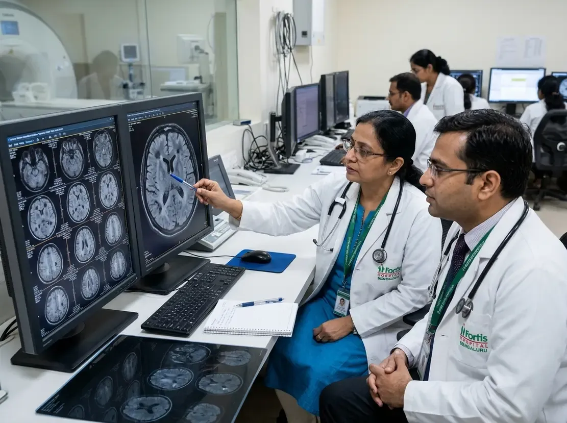
Differentiating multiple sclerosis from its clinical mimics remains a major diagnostic challenge in neuroimmunology. Consequently, the central vein sign has emerged as a crucial imaging biomarker to differentiate demyelinating lesions. However, conventional susceptibility-weighted imaging often fails to show these veins clearly without fluid-attenuated inversion recovery co-registration. Therefore, imaging specialists require refined magnetic resonance sequences to evaluate perivenular lesions with greater diagnostic certainty.
Optimizing the Central Vein Sign on 3T MRI
A recent study evaluated whether an optimized imaging protocol improves lesion visualization at 3 Tesla. Specifically, researchers reduced the flip angle from standard parameters down to five degrees. This protocol adjustment substantially suppressed background parenchymal signal. As a result, the sequence heightened tissue contrast between parenchymal white matter lesions and small central veins. Two independent neuroradiologists evaluated seventeen patients with multiple sclerosis using both standard and modified sequences. Furthermore, the readers assessed the central vein sign blindly while assigning subjective confidence scores to each lesion.
Enhanced Lesion Detection and Reader Confidence
The optimized sequence demonstrated dramatic diagnostic improvements across all evaluated patients. In total, the modified sequence detected 105 positive lesions compared to only 72 lesions on standard imaging. Remarkably, the low-flip-angle protocol identified every single lesion seen on standard imaging plus 33 additional lesions. Additionally, fourteen of the seventeen participants showed higher central vein counts on the optimized sequence. The median number of detected veins increased significantly from 4.0 to 7.0 per patient. Moreover, both radiologists reported significantly higher diagnostic confidence when interpreting the optimized scans. Subject-level classification using the established Select-6 rule also showed superior diagnostic reliability.
Clinical Value for Routine Diagnostic Workflows
Misdiagnosis occurs frequently when clinicians evaluate patients presenting with nonspecific white matter lesions. For instance, disorders like neuromyelitis optica spectrum disorder and small vessel disease can mimic demyelinating plaques. Hence, accurate identification of perivenular venous anatomy helps clinicians confirm or exclude demyelinating disease early. Conventional techniques often require time-consuming co-localization between separate anatomical series. In contrast, this optimized protocol operates effectively as a standalone sequence on standard 3T scanners. Consequently, radiology departments can easily implement this sequence without adding substantial acquisition time or complex post-processing. Clinicians can therefore achieve higher diagnostic precision while minimizing unnecessary therapeutic delays.
Frequently Asked Questions
Q1: What is the central vein sign in multiple sclerosis?
The central vein sign describes a distinct neuroimaging biomarker where a small vein runs centrally through a demyelinating lesion. Furthermore, multiple sclerosis plaques typically form around small parenchymal veins, whereas vascular mimics rarely display this perivenous architecture.
Q2: Why does a lower flip angle improve vein detection?
Reducing the flip angle to five degrees suppresses surrounding brain tissue contrast. Consequently, this adjustment optimizes contrast between paramagnetic deoxyhemoglobin within central veins and surrounding white matter lesions.
Q3: How does the Select-6 rule aid multiple sclerosis diagnosis?
The Select-6 rule is a simplified diagnostic algorithm requiring at least six central vein-positive lesions. Thus, this rule provides an efficient, highly specific threshold that reduces reading time during clinical evaluations.
References
- Hsu YC et al. Improved detection of central vein sign in multiple sclerosis using optimized susceptibility weighted imaging at 3 Tesla. Eur Radiol. 2026 Nov 18. doi: 10.1007/s00330-026-13069-3. PMID: 43049769.
- Sati P, Thomasson D, Li N, et al. Rapid, high-resolution, whole-brain, susceptibility-based MRI of multiple sclerosis. Mult Scler J. 2014;20(11):1464-1470.
- Castellaro M, Tamanti A, Pisani AI, et al. The diagnostic value of the central vein sign in multiple sclerosis: A systematic review and meta-analysis. Mult Scler Relat Disord. 2020;46:102551.