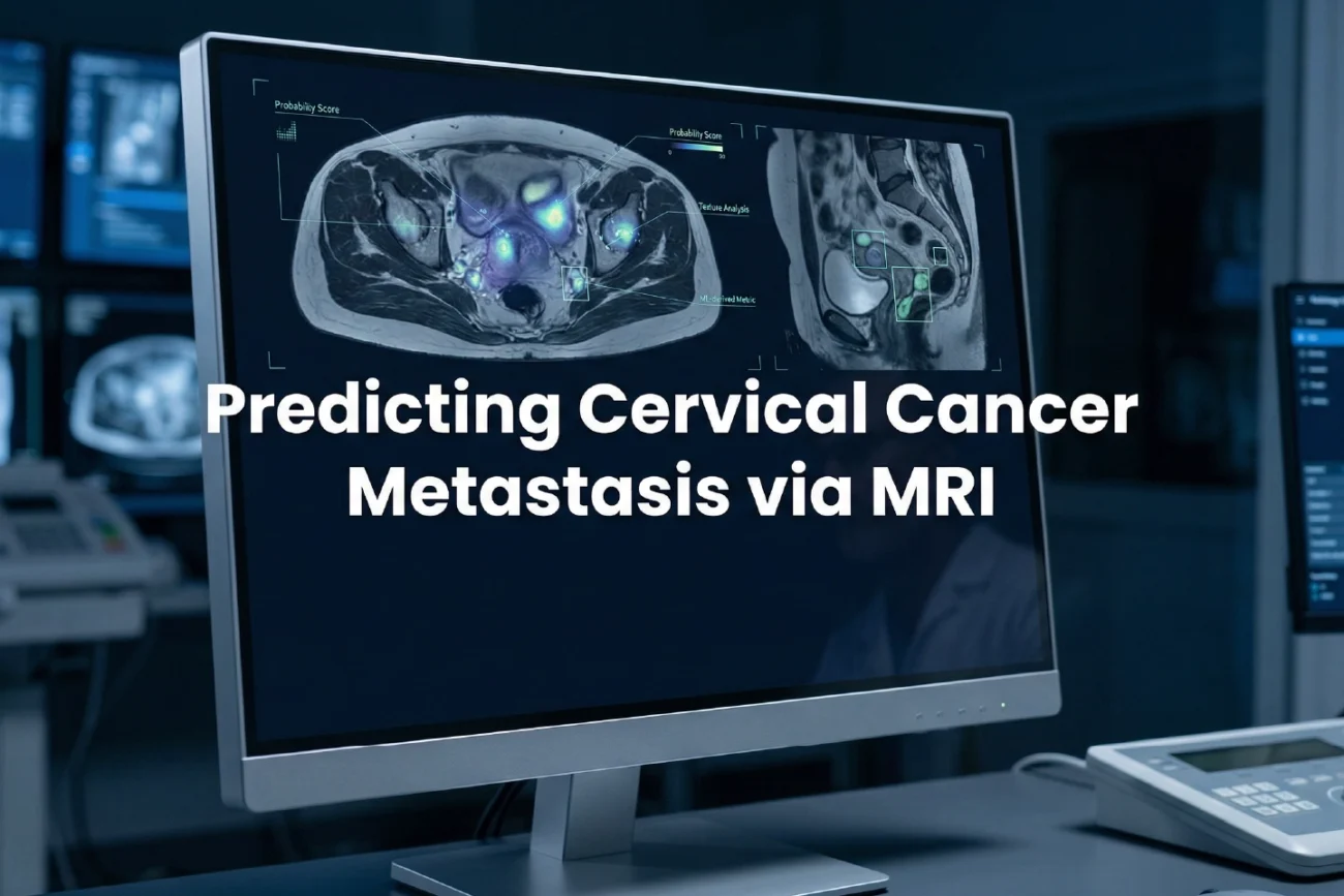
Cervical cancer LNM prediction has traditionally relied on invasive biopsies or post-operative findings. However, a groundbreaking study published in Radiology: Imaging Cancer introduces a superior non-invasive method. This research utilizes time-dependent diffusion MRI (T-dMRI) and macromolecular proton fraction (MPF) mapping. Consequently, doctors can now identify lymph node metastasis (LNM) with unprecedented precision before starting treatment.
Improving Accuracy in Cervical Cancer LNM Prediction
Pre-treatment assessment of lymph nodes is crucial for planning therapy. For instance, LNM-positive patients often require radical surgery and adjuvant chemoradiotherapy. Conversely, LNM-negative patients can avoid the side effects of unnecessary radiation. The researchers discovered that cellularity, intracellular volume fraction, and MPF are significantly higher in metastatic cases. Therefore, these quantitative metrics provide a clearer picture of tumor microstructures than conventional imaging.
Clinical Advantages of Advanced Imaging Metrics
Traditional diffusion-weighted imaging (DWI) often fails to capture fine histopathological details. Furthermore, the new T-dMRI method measures specific parameters like extracellular diffusivity and tumor diameter. When clinicians combine these factors into a diagnostic tool, the results are impressive. Specifically, the area under the curve (AUC) reached 0.95, indicating high reliability. This progress is particularly vital for oncologists in India, where cervical cancer remains a major health concern.
Frequently Asked Questions
Q1: Why is predicting lymph node metastasis important?
Determining nodal status helps clinicians choose between surgery alone or a combination of surgery and radiation. It ensures patients receive the most effective treatment while minimizing long-term complications.
Q2: How does time-dependent MRI differ from standard scans?
Standard MRI focuses on basic tissue morphology and water diffusion. In contrast, time-dependent MRI captures microscopic details such as cell diameter and cellularity, offering a deeper look into the tumor microenvironment.
References
- Meng N et al. Lymph Node Metastases Prediction in Cervical Cancer Using Time-Dependent Diffusion MRI and Macromolecular Proton Fraction Imaging. Radiol Imaging Cancer. 2026 Mar undefined. doi: 10.1148/rycan.250452. PMID: 41758032.
- Li X et al. Clinical value of conventional magnetic resonance imaging combined with diffusion-weighted imaging in predicting pelvic lymph node metastasis of cervical cancer. Front Oncol. 2024.
- Zhang Y et al. Magnetic Resonance Imaging Radiomics-Based Model for Prediction of Lymph Node Metastasis in Cervical Cancer. J Med Imaging Radiat Oncol. 2025.