CT-Guided Core Biopsy Proves Safe for Deep Neck Masses

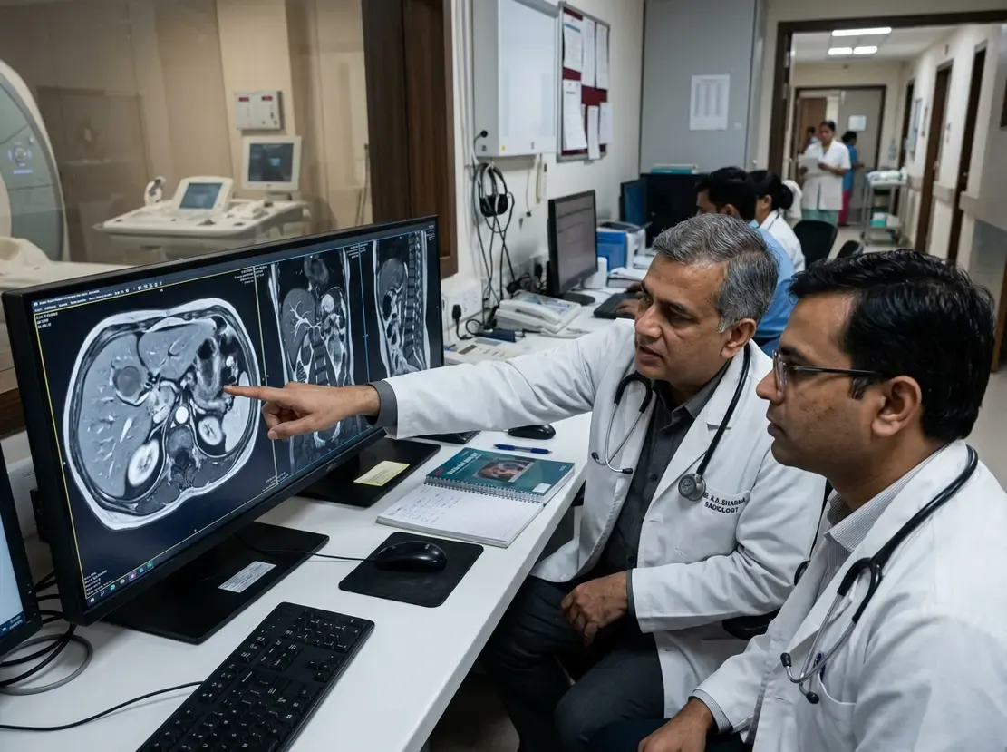
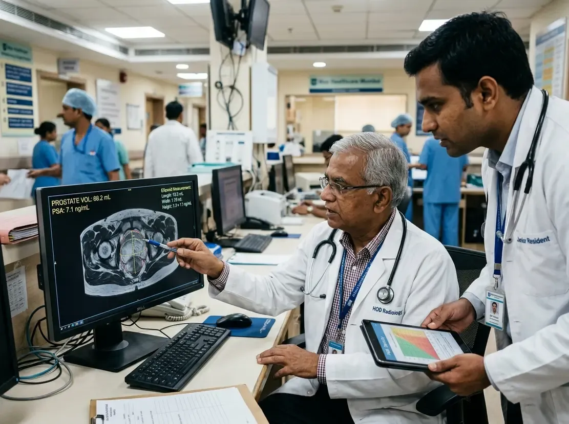
Clinicians frequently face diagnostic hurdles when evaluating lesions in deep cervical compartments. In many patients, overlying mandible and skull base bones obstruct acoustic windows. Consequently, ultrasound cannot reliably guide needles into these anatomical spaces. Recent evidence shows that CT-guided core biopsy provides an effective alternative for such ultrasound-inaccessible lesions. This percutaneous approach yields definitive histological samples while avoiding exploratory surgical morbidity.
Diagnostic Value of CT-Guided Core Biopsy
Researchers conducted a long-term retrospective study evaluating 122 biopsy procedures across 118 patients. Notably, 94.3% of all interventions involved masses located specifically within suprahyoid deep neck spaces. The technique achieved an impressive diagnostic yield of 93.4%. Furthermore, histopathologic concordance between core biopsy specimens and subsequent surgical resections reached 89.5%. Therefore, image guidance under CT offers exceptional diagnostic accuracy for challenging head and neck pathology. In addition, large tissue cores preserve microarchitecture, which facilitates advanced immunohistochemical staining.
Procedural Safety and Clinical Implications
The suprahyoid neck houses critical neurovascular bundles, including the internal carotid artery and jugular vein. However, careful trajectory planning significantly minimizes complication risks. In the analyzed cohort, minor procedure-related complications occurred in only 4.1% of patients. Most importantly, proceduralists reported zero major neurovascular injuries or procedural deaths. Interventional radiologists select tailored trajectories, such as paramaxillary, subzygomatic, or retromandibular pathways. Consequently, patients avoid general anesthesia and the risks associated with open surgical exploration. For oncology teams in India, this minimally invasive method speeds up treatment decisions and reduces healthcare costs.
Frequently Asked Questions
Q1: Why is ultrasound guidance often unfeasible for suprahyoid neck lesions?
Ultrasound waves cannot penetrate intervening cortical bone structures such as the mandible and skull base. Additionally, physiological air spaces in the pharynx create acoustic barriers, obscuring deep needle visualization.
Q2: What is the typical complication rate associated with CT-guided core biopsy in deep neck spaces?
Clinical studies report low complication rates around 4%, which primarily consist of self-limiting minor hematomas. Furthermore, serious complications like major vascular laceration remain exceedingly rare with precise planning.
Q3: How does core needle biopsy compare to fine-needle aspiration in this setting?
Core needle biopsy provides intact tissue cylinders that preserve architectural detail for histopathology. Consequently, it allows comprehensive immunohistochemistry and molecular profiling, whereas fine-needle aspiration often yields limited cellular material.
References
- Kim J et al. CT-guided core needle biopsy in ultrasound-inaccessible suprahyoid deep neck spaces. Eur Radiol. 2026 Sep 28. doi: 10.1007/s00330-026-12867-z. PMID: 42803938.
- McKnight CD, Glastonbury CM, Ibrahim M, Rivas-Rodriguez F, Srinivasan A. Techniques and Approaches for Safe, High-Yield CT-Guided Suprahyoid Head and Neck Biopsies. AJR Am J Roentgenol. 2017;208(5):917-922. doi: 10.2214/AJR.16.16558.
- Jordanov NP, et al. Percutaneous CT-Guided Core Needle Biopsies of Head and Neck Masses: Review of 184 Cases at a Single Academic Institution, Common and Special Techniques, Diagnostic Yield, and Safety. AJNR Am J Neuroradiol. 2022;43(1):118-125. doi: 10.3174/ajnr.A7366.