
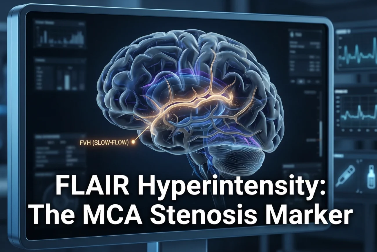
Recent research identifies FLAIR vascular hyperintensity as a primary indicator of hemodynamic impairment in patients with middle cerebral artery (MCA) stenosis. Specifically, this radiographic sign represents slow blood flow within leptomeningeal collaterals. Therefore, it serves as a critical biomarker for assessing stroke risk and vascular burden. Consequently, clinicians must recognize its presence to optimize patient management and treatment strategies.
Pathophysiology of FLAIR Vascular Hyperintensity
The \”hyperintense vessel sign\” typically indicates stagnant flow or stasis in distal arteries. Notably, severe stenosis often triggers this phenomenon by reducing perfusion pressure in the brain. However, the presence of these hyperintensities does not always mean an infarction has occurred. Instead, it frequently highlights tissue at risk of future ischemic damage. Thus, detecting these signals early remains essential for effective neuroprotection.
Vascular Lesion Burden and Enhancement
Researchers recently explored how vascular wall characteristics influence these imaging markers. Specifically, marked enhancement of the vessel wall strongly correlates with increased hyperintensity burden. Furthermore, infarction and severe stenosis show independent associations with the presence of these signs. Moreover, the study utilized multivariate regression to confirm these links. Additionally, these findings emphasize the complex relationship between vessel wall health and distal hemodynamics.
Diagnostic Performance in Clinical Practice
The study demonstrated that combining clinical and imaging factors improves diagnostic accuracy. Specifically, the integrated model achieved an area under the curve (AUC) of 0.814. Furthermore, this approach yielded a sensitivity of 77.9% and a specificity of 73.7%. Notably, these metrics highlight the model’s ability to discriminate between high and low vascular burdens. Therefore, such tools offer significant value in routine clinical practice for stroke specialists.
Frequently Asked Questions
Q1: What does FLAIR vascular hyperintensity represent?
It represents slow or stagnant blood flow, typically in the leptomeningeal collateral vessels of the brain.
Q2: How does it help in stroke risk assessment?
Its presence indicates severe hemodynamic impairment, identifying patients who may require aggressive revascularization strategies to prevent infarction.
Q3: Which factors are most associated with this marker?
Severe arterial stenosis, established infarction, and marked vessel wall enhancement show the strongest independent links to this imaging sign.
References
- Jiang C et al. FLAIR vascular hyperintensity: association with infarction and vascular lesion burden in patients with middle cerebral artery stenosis. Eur Radiol. 2026 Mar 12. doi: 10.1007/s00330-025-12282-w. PMID: 41817708.
- Shin NY et al. Fluid-Attenuated Inversion Recovery Vascular Hyperintensity in Cerebrovascular Disease: A Review for Radiologists and Clinicians. Front Neurol. 2021;12:791090.
- Sanossian N et al. Significance of hyperintense vessels on FLAIR MRI in acute stroke. Neurology. 2009;72(14):1240-5.